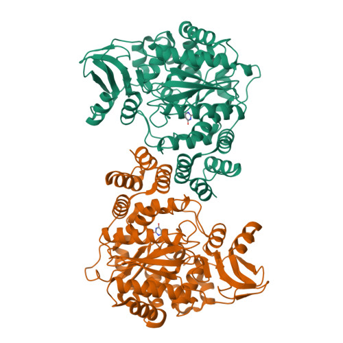
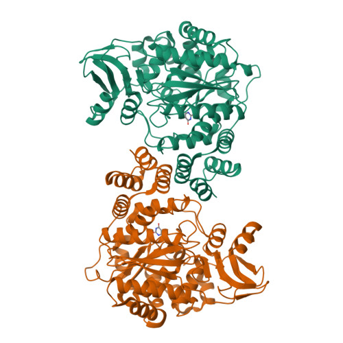
2WK6

Structural features of native human thymidine phosphorylase and in complex with 5-iodouracil


Experimental Data Snapshot

  • Method: X-RAY DIFFRACTION
  • Resolution: 2.50 Å
  • R-Value Free: 0.283 
  • R-Value Work: 0.227 
  • R-Value Observed: 0.227 

Starting Model: experimental
View more details

wwPDB Validation   3D Report Full Report


Ligand Structure Quality Assessment 


This is version 1.4 of the entry. See complete history


Literature

Structures of Native Human Thymidine Phosphorylase and in Complex with 5-Iodouracil.

Mitsiki, E.Papageorgiou, A.C.Iyer, S.Thiyagarajan, N.Prior, S.H.Sleep, D.Finnis, C.Acharya, K.R.

(2009) Biochem Biophys Res Commun 386: 666

  • DOI: https://doi.org/10.1016/j.bbrc.2009.06.104
  • Primary Citation of Related Structures:  
    2WK5, 2WK6

  • PubMed Abstract: 

    Thymidine phosphorylase (TP) first identified as platelet derived endothelial cell growth factor (PD-ECGF) plays a key role in nucleoside metabolism. Human TP (hTP) is implicated in angiogenesis and is overexpressed in several solid tumors. Here, we report the crystal structures of recombinant hTP and its complex with a substrate 5-iodouracil (5IUR) at 3.0 and 2.5A, respectively. In addition, we provide information on the role of specific residues in the enzymatic activity of hTP through mutagenesis and kinetic studies.


  • Organizational Affiliation

    Department of Biology and Biochemistry, University of Bath, Claverton Down, Bath BA2 7AY, UK.


Macromolecules
Find similar proteins by:  (by identity cutoff)  |  3D Structure
Entity ID: 1
MoleculeChains Sequence LengthOrganismDetailsImage
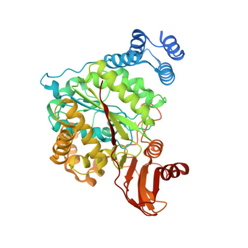
THYMIDINE PHOSPHORYLASE
A, B
482Homo sapiensMutation(s): 0 
EC: 2.4.2.4
UniProt & NIH Common Fund Data Resources
Find proteins for P19971 (Homo sapiens)
Explore P19971 
Go to UniProtKB:  P19971
PHAROS:  P19971
GTEx:  ENSG00000025708 
Entity Groups  
Sequence Clusters30% Identity50% Identity70% Identity90% Identity95% Identity100% Identity
UniProt GroupP19971
Sequence Annotations
Expand
  • Reference Sequence
Small Molecules
Binding Affinity Annotations 
IDSourceBinding Affinity
IUR PDBBind:  2WK6 Ki: 4.80e+5 (nM) from 1 assay(s)
Experimental Data & Validation

Experimental Data

  • Method: X-RAY DIFFRACTION
  • Resolution: 2.50 Å
  • R-Value Free: 0.283 
  • R-Value Work: 0.227 
  • R-Value Observed: 0.227 
  • Space Group: P 21 21 21
Unit Cell:
Length ( Å )Angle ( ˚ )
a = 61.911α = 90
b = 67.266β = 90
c = 212.274γ = 90
Software Package:
Software NamePurpose
CNSrefinement
DENZOdata reduction
SCALEPACKdata scaling
MOLREPphasing

Structure Validation

View Full Validation Report


Ligand Structure Quality Assessment 


Entry History 

Deposition Data

Revision History  (Full details and data files)

  • Version 1.0: 2009-07-07
    Type: Initial release
  • Version 1.1: 2011-05-08
    Changes: Version format compliance
  • Version 1.2: 2011-07-13
    Changes: Version format compliance
  • Version 1.3: 2018-01-17
    Changes: Data collection
  • Version 1.4: 2023-12-13
    Changes: Data collection, Database references, Derived calculations, Other, Refinement description