2X10 | pdb_00002x10

Crystal structure of the complete EphA2 ectodomain


Experimental Data Snapshot

  • Method: X-RAY DIFFRACTION
  • Resolution: 3.00 Å
  • R-Value Free: 
    0.299 (Depositor), 0.290 (DCC) 
  • R-Value Work: 
    0.236 (Depositor), 0.230 (DCC) 
  • R-Value Observed: 
    0.239 (Depositor) 

Starting Models: experimental
View more details

wwPDB Validation   3D Report Full Report


Ligand Structure Quality Assessment 

Created with Raphaël 2.3.0Worse 01 BetterLigand structure goodness of fit to experimental dataBest fitted NAGClick on this verticalbar to view details

This is version 1.5 of the entry. See complete history


Literature

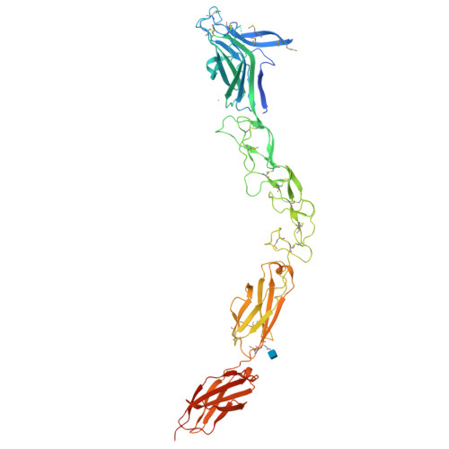
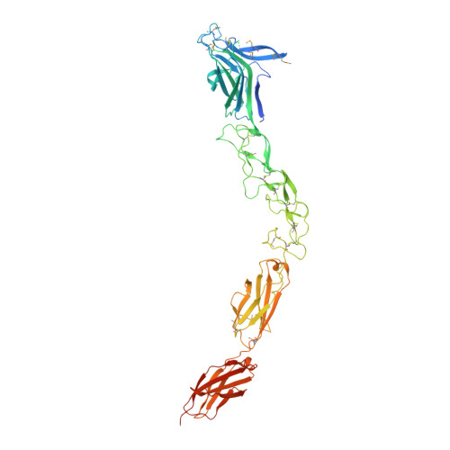
An Extracellular Steric Seeding Mechanism for Eph-Ephrin Signalling Platform Assembly

Seiradake, E.Harlos, K.Sutton, G.Aricescu, A.R.Jones, E.Y.

(2010) Nat Struct Mol Biol 17: 398

  • DOI: https://doi.org/10.1038/nsmb.1782
  • Primary Citation of Related Structures:  
    2X10, 2X11

  • PubMed Abstract: 

    Erythropoetin-producing hepatoma (Eph) receptors are cell-surface protein tyrosine kinases mediating cell-cell communication. Upon activation, they form signaling clusters. We report crystal structures of the full ectodomain of human EphA2 (eEphA2) both alone and in complex with the receptor-binding domain of the ligand ephrinA5 (ephrinA5 RBD). Unliganded eEphA2 forms linear arrays of staggered parallel receptors involving two patches of residues conserved across A-class Ephs. eEphA2-ephrinA5 RBD forms a more elaborate assembly, whose interfaces include the same conserved regions on eEphA2, but rearranged to accommodate ephrinA5 RBD. Cell-surface expression of mutant EphA2s showed that these interfaces are critical for localization at cell-cell contacts and activation-dependent degradation. Our results suggest a 'nucleation' mechanism whereby a limited number of ligand-receptor interactions 'seed' an arrangement of receptors which can propagate into extended signaling arrays.


  • Organizational Affiliation

    Division of Structural Biology, Wellcome Trust Centre for Human Genetics, University of Oxford, Oxford, UK.


Macromolecules
Find similar proteins by:  (by identity cutoff)  |  3D Structure
Entity ID: 1
MoleculeChains Sequence LengthOrganismDetailsImage
EPHRIN TYPE-A RECEPTOR 2545Homo sapiensMutation(s): 0 
EC: 2.7.10.1
UniProt & NIH Common Fund Data Resources
Find proteins for P29317 (Homo sapiens)
Explore P29317 
Go to UniProtKB:  P29317
PHAROS:  P29317
GTEx:  ENSG00000142627 
Entity Groups  
Sequence Clusters30% Identity50% Identity70% Identity90% Identity95% Identity100% Identity
UniProt GroupP29317
Glycosylation
Glycosylation Sites: 1Go to GlyGen: P29317-1
Sequence Annotations
Expand
  • Reference Sequence
Small Molecules
Modified Residues  1 Unique
IDChains TypeFormula2D DiagramParent
MSE
Query on MSE
A
L-PEPTIDE LINKINGC5 H11 N O2 SeMET
Experimental Data & Validation

Experimental Data

  • Method: X-RAY DIFFRACTION
  • Resolution: 3.00 Å
  • R-Value Free:  0.299 (Depositor), 0.290 (DCC) 
  • R-Value Work:  0.236 (Depositor), 0.230 (DCC) 
  • R-Value Observed: 0.239 (Depositor) 
Space Group: P 1
Unit Cell:
Length ( Å )Angle ( ˚ )
a = 41.92α = 93.02
b = 44.65β = 98.22
c = 93.42γ = 112.12
Software Package:
Software NamePurpose
PHENIXrefinement
XDSdata reduction
MOSFLMdata reduction
XSCALEdata scaling
PHASERphasing

Structure Validation

View Full Validation Report


Ligand Structure Quality Assessment 

Created with Raphaël 2.3.0Worse 01 BetterLigand structure goodness of fit to experimental dataBest fitted NAGClick on this verticalbar to view details

Entry History 

Deposition Data

Revision History  (Full details and data files)

  • Version 1.0: 2010-03-16
    Type: Initial release
  • Version 1.1: 2013-01-30
    Changes: Advisory, Atomic model, Database references, Derived calculations, Non-polymer description, Other, Refinement description, Version format compliance
  • Version 1.2: 2018-02-28
    Changes: Source and taxonomy
  • Version 1.3: 2020-07-29
    Type: Remediation
    Reason: Carbohydrate remediation
    Changes: Advisory, Data collection, Derived calculations, Other, Structure summary
  • Version 1.4: 2023-12-20
    Changes: Data collection, Database references, Refinement description, Structure summary
  • Version 1.5: 2024-11-06
    Changes: Structure summary