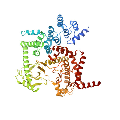
2X6K

THE CRYSTAL STRUCTURE OF THE DROSOPHILA CLASS III PI3-KINASE VPS34 IN COMPLEX WITH PI-103


Experimental Data Snapshot

  • Method: X-RAY DIFFRACTION
  • Resolution: 3.50 Å
  • R-Value Free: 0.296 
  • R-Value Work: 0.235 
  • R-Value Observed: 0.238 

Starting Model: experimental
View more details

wwPDB Validation   3D Report Full Report


Ligand Structure Quality Assessment 


This is version 1.3 of the entry. See complete history


Literature

Shaping Development of Autophagy Inhibitors with the Structure of the Lipid Kinase Vps34.

Miller, S.Tavshanjian, B.Oleksy, A.Perisic, O.Houseman, B.T.Shokat, K.M.Williams, R.L.

(2010) Science 327: 1638

  • DOI: https://doi.org/10.1126/science.1184429
  • Primary Citation of Related Structures:  
    2X6F, 2X6H, 2X6I, 2X6J, 2X6K

  • PubMed Abstract: 

    Phosphoinositide 3-kinases (PI3Ks) are lipid kinases with diverse roles in health and disease. The primordial PI3K, Vps34, is present in all eukaryotes and has essential roles in autophagy, membrane trafficking, and cell signaling. We solved the crystal structure of Vps34 at 2.9 angstrom resolution, which revealed a constricted adenine-binding pocket, suggesting the reason that specific inhibitors of this class of PI3K have proven elusive. Both the phosphoinositide-binding loop and the carboxyl-terminal helix of Vps34 mediate catalysis on membranes and suppress futile adenosine triphosphatase cycles. Vps34 appears to alternate between a closed cytosolic form and an open form on the membrane. Structures of Vps34 complexes with a series of inhibitors reveal the reason that an autophagy inhibitor preferentially inhibits Vps34 and underpin the development of new potent and specific Vps34 inhibitors.


  • Organizational Affiliation

    Medical Research Council Laboratory of Molecular Biology, Hills Road, Cambridge CB2 0QH, UK.


Macromolecules
Find similar proteins by:  (by identity cutoff)  |  3D Structure
Entity ID: 1
MoleculeChains Sequence LengthOrganismDetailsImage
PHOSPHOTIDYLINOSITOL 3 KINASE 59F
A, B
696Drosophila melanogasterMutation(s): 1 
EC: 2.7.1.137 (PDB Primary Data), 2.7.1.153 (PDB Primary Data), 2.7.1.154 (PDB Primary Data)
UniProt
Find proteins for Q9W1M7 (Drosophila melanogaster)
Explore Q9W1M7 
Go to UniProtKB:  Q9W1M7
Entity Groups  
Sequence Clusters30% Identity50% Identity70% Identity90% Identity95% Identity100% Identity
UniProt GroupQ9W1M7
Sequence Annotations
Expand
  • Reference Sequence
Experimental Data & Validation

Experimental Data

  • Method: X-RAY DIFFRACTION
  • Resolution: 3.50 Å
  • R-Value Free: 0.296 
  • R-Value Work: 0.235 
  • R-Value Observed: 0.238 
  • Space Group: I 21 21 21
Unit Cell:
Length ( Å )Angle ( ˚ )
a = 111.44α = 90
b = 155.68β = 90
c = 244γ = 90
Software Package:
Software NamePurpose
REFMACrefinement
iMOSFLMdata reduction
SCALAdata scaling
PHASERphasing

Structure Validation

View Full Validation Report


Ligand Structure Quality Assessment 


Entry History 

Deposition Data

Revision History  (Full details and data files)

  • Version 1.0: 2010-04-07
    Type: Initial release
  • Version 1.1: 2011-07-13
    Changes: Advisory, Refinement description, Version format compliance
  • Version 1.2: 2018-01-24
    Changes: Source and taxonomy
  • Version 1.3: 2023-12-20
    Changes: Data collection, Database references, Derived calculations, Other, Refinement description