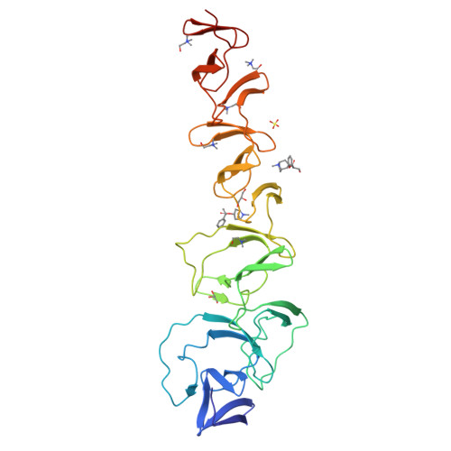
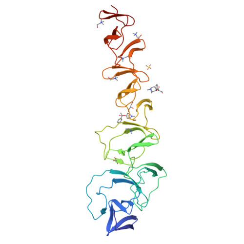
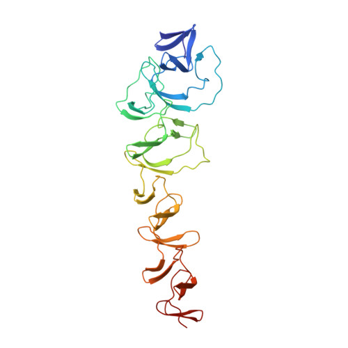
2X8P

Crystal Structure of CbpF in Complex with Atropine by Co- Crystallization


Experimental Data Snapshot

  • Method: X-RAY DIFFRACTION
  • Resolution: 2.27 Å
  • R-Value Free: 0.288 
  • R-Value Work: 0.226 
  • R-Value Observed: 0.226 

Starting Model: experimental
View more details

wwPDB Validation   3D Report Full Report


Ligand Structure Quality Assessment 


This is version 1.3 of the entry. See complete history


Literature

Crystal Structures of Cbpf Complexed with Atropine and Ipratropium Reveal Clues for the Design of Novel Antimicrobials Against Streptococcus Pneumoniae.

Silva-Martin, N.Retamosa, M.G.Maestro, B.Bartual, S.G.Rodes, M.J.Garcia, P.Sanz, J.M.Hermoso, J.A.

(2013) Biochim Biophys Acta 1840: 129

  • DOI: https://doi.org/10.1016/j.bbagen.2013.09.006
  • Primary Citation of Related Structures:  
    2X8M, 2X8P

  • PubMed Abstract: 

    Streptococcus pneumoniae is a major pathogen responsible of important diseases worldwide such as pneumonia and meningitis. An increasing resistance level hampers the use of currently available antibiotics to treat pneumococcal diseases. Consequently, it is desirable to find new targets for the development of novel antimicrobial drugs to treat pneumococcal infections. Surface choline-binding proteins (CBPs) are essential in bacterial physiology and infectivity. In this sense, esters of bicyclic amines (EBAs) such as atropine and ipratropium have been previously described to act as choline analogs and effectively compete with teichoic acids on binding to CBPs, consequently preventing in vitro pneumococcal growth, altering cell morphology and reducing cell viability. With the aim of gaining a deeper insight into the structural determinants of the strong interaction between CBPs and EBAs, the three-dimensional structures of choline-binding protein F (CbpF), one of the most abundant proteins in the pneumococcal cell wall, complexed with atropine and ipratropium, have been obtained. The choline analogs bound both to the carboxy-terminal module, involved in cell wall binding, and, unexpectedly, also to the amino-terminal module, that possesses a regulatory role in pneumococcal autolysis. Analysis of the complexes confirmed the importance of the tropic acid moiety of the EBAs on the strength of the binding, through π-π interactions with aromatic residues in the binding site. These results represent the first example describing the molecular basis of the inhibition of CBPs by EBA molecules and pave the way for the development of new generations of antipneumococcal drugs.


  • Organizational Affiliation

    Departamento de Cristalografía y Biología Estructural, Instituto de Química-Física 'Rocasolano', Consejo Superior de Investigaciones Científicas, Serrano 119, 28006 Madrid, Spain.


Macromolecules
Find similar proteins by:  (by identity cutoff)  |  3D Structure
Entity ID: 1
MoleculeChains Sequence LengthOrganismDetailsImage
CHOLINE-BINDING PROTEIN F311Streptococcus pneumoniae R6Mutation(s): 0 
UniProt
Find proteins for Q8DR52 (Streptococcus pneumoniae (strain ATCC BAA-255 / R6))
Explore Q8DR52 
Go to UniProtKB:  Q8DR52
Entity Groups  
Sequence Clusters30% Identity50% Identity70% Identity90% Identity95% Identity100% Identity
UniProt GroupQ8DR52
Sequence Annotations
Expand
  • Reference Sequence
Small Molecules
Ligands 4 Unique
IDChains Name / Formula / InChI Key2D Diagram3D Interactions
OIN
Query on OIN

Download Ideal Coordinates CCD File 
C [auth A],
E [auth A]
(1R,5S)-8-METHYL-8-AZABICYCLO[3.2.1]OCT-3-YL (2R)-3-HYDROXY-2-PHENYLPROPANOATE
C17 H23 N O3
RKUNBYITZUJHSG-QKPAOTATSA-N
CHT
Query on CHT

Download Ideal Coordinates CCD File 
G [auth A],
H [auth A],
I [auth A],
J [auth A],
K [auth A]
CHOLINE ION
C5 H14 N O
OEYIOHPDSNJKLS-UHFFFAOYSA-N
SO4
Query on SO4

Download Ideal Coordinates CCD File 
B [auth A]SULFATE ION
O4 S
QAOWNCQODCNURD-UHFFFAOYSA-L
GOL
Query on GOL

Download Ideal Coordinates CCD File 
D [auth A],
F [auth A]
GLYCEROL
C3 H8 O3
PEDCQBHIVMGVHV-UHFFFAOYSA-N
Experimental Data & Validation

Experimental Data

  • Method: X-RAY DIFFRACTION
  • Resolution: 2.27 Å
  • R-Value Free: 0.288 
  • R-Value Work: 0.226 
  • R-Value Observed: 0.226 
  • Space Group: P 21 21 2
Unit Cell:
Length ( Å )Angle ( ˚ )
a = 52.54α = 90
b = 116.077β = 90
c = 73.165γ = 90
Software Package:
Software NamePurpose
CNSrefinement
MOSFLMdata reduction
SCALEPACKdata scaling
MOLREPphasing

Structure Validation

View Full Validation Report


Ligand Structure Quality Assessment 


Entry History 

Deposition Data

Revision History  (Full details and data files)

  • Version 1.0: 2011-04-06
    Type: Initial release
  • Version 1.1: 2013-10-02
    Changes: Database references, Non-polymer description, Refinement description, Version format compliance
  • Version 1.2: 2013-11-13
    Changes: Database references
  • Version 1.3: 2023-12-20
    Changes: Data collection, Database references, Derived calculations, Other, Refinement description