2XAH

Crystal structure of LSD1-CoREST in complex with (+)-trans-2- phenylcyclopropyl-1-amine


Experimental Data Snapshot

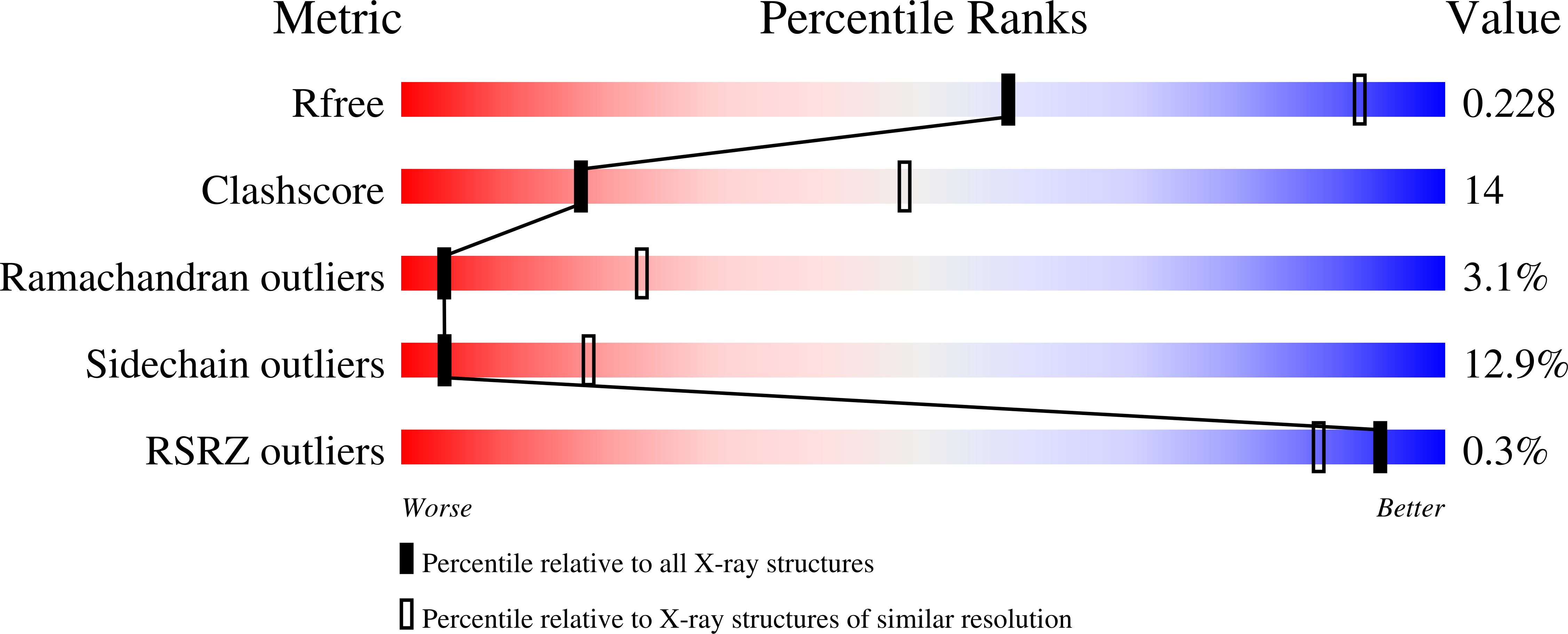
  • Method: X-RAY DIFFRACTION
  • Resolution: 3.10 Å
  • R-Value Free: 0.228 
  • R-Value Work: 0.208 
  • R-Value Observed: 0.209 

wwPDB Validation   3D Report Full Report


Ligand Structure Quality Assessment 


This is version 1.2 of the entry. See complete history


Literature

Biochemical, Structural, and Biological Evaluation of Tranylcypromine Derivatives as Inhibitors of Histone Demethylases Lsd1 and Lsd2.

Binda, C.Valente, S.Romanenghi, M.Pilotto, S.Cirilli, R.Karytinos, A.Ciossani, G.Botrugno, O.A.Forneris, F.Tardugno, M.Edmondson, D.E.Minucci, S.Mattevi, A.Mai, A.

(2010) J Am Chem Soc 132: 6827

  • DOI: https://doi.org/10.1021/ja101557k
  • Primary Citation of Related Structures:  
    2XAF, 2XAG, 2XAH, 2XAJ, 2XAQ, 2XAS

  • PubMed Abstract: 

    LSD1 and LSD2 histone demethylases are implicated in a number of physiological and pathological processes, ranging from tumorigenesis to herpes virus infection. A comprehensive structural, biochemical, and cellular study is presented here to probe the potential of these enzymes for epigenetic therapies. This approach employs tranylcypromine as a chemical scaffold for the design of novel demethylase inhibitors. This drug is a clinically validated antidepressant known to target monoamine oxidases A and B. These two flavoenzymes are structurally related to LSD1 and LSD2. Mechanistic and crystallographic studies of tranylcypromine inhibition reveal a lack of selectivity and differing covalent modifications of the FAD cofactor depending on the enantiomeric form. These findings are pharmacologically relevant, since tranylcypromine is currently administered as a racemic mixture. A large set of tranylcypromine analogues were synthesized and screened for inhibitory activities. We found that the common evolutionary origin of LSD and MAO enzymes, despite their unrelated functions and substrate specificities, is reflected in related ligand-binding properties. A few compounds with partial enzyme selectivity were identified. The biological activity of one of these new inhibitors was evaluated with a cellular model of acute promyelocytic leukemia chosen since its pathogenesis includes aberrant activities of several chromatin modifiers. Marked effects on cell differentiation and an unprecedented synergistic activity with antileukemia drugs were observed. These data demonstrate that these LSD1/2 inhibitors are of potential relevance for the treatment of promyelocytic leukemia and, more generally, as tools to alter chromatin state with promise of a block of tumor progression.


  • Organizational Affiliation

    Department of Genetics and Microbiology, University of Pavia, Via Ferrata 1, 27100 Pavia, Italy.


Macromolecules
Find similar proteins by:  (by identity cutoff)  |  3D Structure
Entity ID: 1
MoleculeChains Sequence LengthOrganismDetailsImage
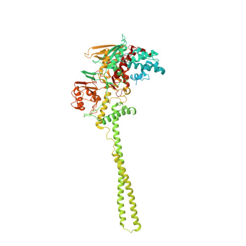
LYSINE-SPECIFIC HISTONE DEMETHYLASE 1852Homo sapiensMutation(s): 0 
EC: 1 (PDB Primary Data), 1.14.99.66 (UniProt)
UniProt & NIH Common Fund Data Resources
Find proteins for O60341 (Homo sapiens)
Explore O60341 
Go to UniProtKB:  O60341
PHAROS:  O60341
GTEx:  ENSG00000004487 
Entity Groups  
Sequence Clusters30% Identity50% Identity70% Identity90% Identity95% Identity100% Identity
UniProt GroupO60341
Sequence Annotations
Expand
  • Reference Sequence
Find similar proteins by:  (by identity cutoff)  |  3D Structure
Entity ID: 2
MoleculeChains Sequence LengthOrganismDetailsImage
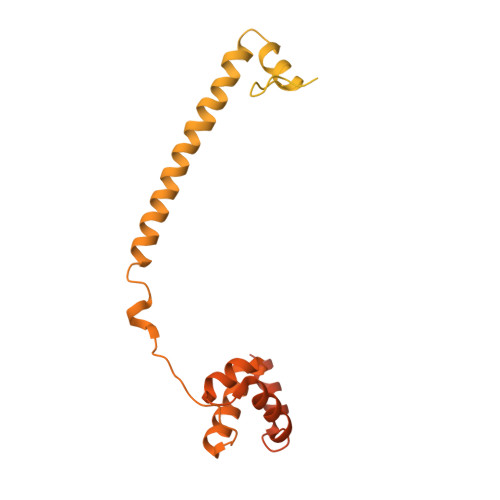
REST COREPRESSOR 1482Homo sapiensMutation(s): 0 
UniProt & NIH Common Fund Data Resources
Find proteins for Q9UKL0 (Homo sapiens)
Explore Q9UKL0 
Go to UniProtKB:  Q9UKL0
PHAROS:  Q9UKL0
GTEx:  ENSG00000089902 
Entity Groups  
Sequence Clusters30% Identity50% Identity70% Identity90% Identity95% Identity100% Identity
UniProt GroupQ9UKL0
Sequence Annotations
Expand
  • Reference Sequence
Small Molecules
Binding Affinity Annotations 
IDSourceBinding Affinity
3PL PDBBind:  2XAH Ki: 2.84e+5 (nM) from 1 assay(s)
Experimental Data & Validation

Experimental Data

  • Method: X-RAY DIFFRACTION
  • Resolution: 3.10 Å
  • R-Value Free: 0.228 
  • R-Value Work: 0.208 
  • R-Value Observed: 0.209 
  • Space Group: I 2 2 2
Unit Cell:
Length ( Å )Angle ( ˚ )
a = 119.959α = 90
b = 181.498β = 90
c = 234.706γ = 90
Software Package:
Software NamePurpose
REFMACrefinement
MOSFLMdata reduction
SCALAdata scaling

Structure Validation

View Full Validation Report


Ligand Structure Quality Assessment 


Entry History 

Revision History  (Full details and data files)

  • Version 1.0: 2010-05-05
    Type: Initial release
  • Version 1.1: 2011-10-12
    Changes: Database references, Derived calculations, Refinement description, Version format compliance
  • Version 1.2: 2024-05-08
    Changes: Data collection, Database references, Derived calculations, Other