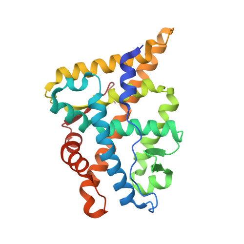
2YLO

TARGETING THE BINDING FUNCTION 3 SITE OF THE ANDROGEN RECEPTOR THROUGH IN SILICO MOLECULAR MODELING


Experimental Data Snapshot

  • Method: X-RAY DIFFRACTION
  • Resolution: 2.50 Å
  • R-Value Free: 0.289 
  • R-Value Work: 0.224 
  • R-Value Observed: 0.227 

Starting Model: experimental
View more details

wwPDB Validation   3D Report Full Report


Ligand Structure Quality Assessment 


This is version 1.3 of the entry. See complete history


Literature

Targeting the Binding Function 3 (Bf3) Site of the Human Androgen Receptor Through Virtual Screening.

Lack, N.A.Axerio-Cilies, P.Tavassoli, P.Han, F.Q.Chan, K.H.Feau, C.Leblanc, E.Guns, E.T.Guy, R.K.Rennie, P.S.Cherkasov, A.

(2011) J Med Chem 54: 8563

  • DOI: https://doi.org/10.1021/jm201098n
  • Primary Citation of Related Structures:  
    2YLO, 2YLP, 2YLQ, 3ZQT

  • PubMed Abstract: 

    The androgen receptor (AR) is the best studied drug target for the treatment of prostate cancer. While there are a number of drugs that target the AR, they all work through the same mechanism of action and are prone to the development of drug resistance. There is a large unmet need for novel AR inhibitors which work through alternative mechanism(s). Recent studies have identified a novel site on the AR called binding function 3 (BF3) that is involved into AR transcriptional activity. In order to identify inhibitors that target the BF3 site, we have conducted a large-scale in silico screen followed by experimental evaluation. A number of compounds were identified that effectively inhibited the AR transcriptional activity with no obvious cytotoxicity. The mechanism of action of these compounds was validated by biochemical assays and X-ray crystallography. These findings lay a foundation for the development of alternative or supplementary therapies capable of combating prostate cancer even in its antiandrogen resistant forms.


  • Organizational Affiliation

    Vancouver Prostate Centre, University of British Columbia, 2660 Oak Street, Vancouver, British Columbia V6H 3Z6, Canada.


Macromolecules
Find similar proteins by:  (by identity cutoff)  |  3D Structure
Entity ID: 1
MoleculeChains Sequence LengthOrganismDetailsImage
ANDROGEN RECEPTOR256Homo sapiensMutation(s): 0 
UniProt & NIH Common Fund Data Resources
Find proteins for P10275 (Homo sapiens)
Explore P10275 
Go to UniProtKB:  P10275
PHAROS:  P10275
GTEx:  ENSG00000169083 
Entity Groups  
Sequence Clusters30% Identity50% Identity70% Identity90% Identity95% Identity100% Identity
UniProt GroupP10275
Sequence Annotations
Expand
  • Reference Sequence
Small Molecules
Binding Affinity Annotations 
IDSourceBinding Affinity
TES BindingDB:  2YLO Ki: min: 1.4, max: 29 (nM) from 5 assay(s)
Kd: 0.4 (nM) from 1 assay(s)
IC50: min: 2.7, max: 3090.3 (nM) from 11 assay(s)
EC50: min: 1.1, max: 3.16 (nM) from 4 assay(s)
YLO BindingDB:  2YLO IC50: min: 1.00e+4, max: 5.00e+4 (nM) from 3 assay(s)
PDBBind:  2YLO IC50: 1.31e+4 (nM) from 1 assay(s)
Experimental Data & Validation

Experimental Data

  • Method: X-RAY DIFFRACTION
  • Resolution: 2.50 Å
  • R-Value Free: 0.289 
  • R-Value Work: 0.224 
  • R-Value Observed: 0.227 
  • Space Group: P 21 21 21
Unit Cell:
Length ( Å )Angle ( ˚ )
a = 55.32α = 90
b = 66.5β = 90
c = 73.12γ = 90
Software Package:
Software NamePurpose
REFMACrefinement
MOSFLMdata reduction
MOSFLMdata scaling
PHASERphasing

Structure Validation

View Full Validation Report


Ligand Structure Quality Assessment 


Entry History 

Revision History  (Full details and data files)

  • Version 1.0: 2011-07-06
    Type: Initial release
  • Version 1.1: 2011-12-07
    Changes: Database references, Version format compliance
  • Version 1.2: 2012-01-11
    Changes: Other
  • Version 1.3: 2023-12-20
    Changes: Data collection, Database references, Derived calculations, Other, Refinement description