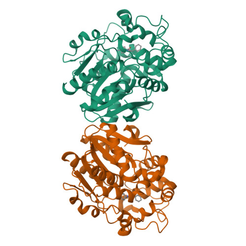
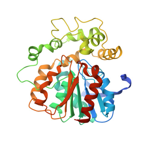
3ANS

Human soluble epoxide hydrolase in complex with a synthetic inhibitor


Experimental Data Snapshot

  • Method: X-RAY DIFFRACTION
  • Resolution: 1.98 Å
  • R-Value Free: 0.260 
  • R-Value Work: 0.219 
  • R-Value Observed: 0.221 

Starting Model: experimental
View more details

wwPDB Validation   3D Report Full Report


Ligand Structure Quality Assessment 


This is version 1.4 of the entry. See complete history


Literature

A practical use of ligand efficiency indices out of the fragment-based approach: ligand efficiency-guided lead identification of soluble epoxide hydrolase inhibitors

Tanaka, D.Tsuda, Y.Shiyama, T.Nishimura, T.Chiyo, N.Tominaga, Y.Sawada, N.Mimoto, T.Kusunose, N.

(2011) J Med Chem 54: 851-857

  • DOI: https://doi.org/10.1021/jm101273e
  • Primary Citation of Related Structures:  
    3ANS, 3ANT

  • PubMed Abstract: 

    Ligand efficiency is frequently used to evaluate fragment compounds in fragment-based drug discovery. We applied ligand efficiency indices in a conventional virtual screening-initiated lead generation study of soluble epoxide hydrolase inhibitors. From a considerable number of screening hits, we carefully selected a compound exhibiting relatively weak inhibitory activity but high ligand efficiency. This ligand efficiency-guided selection could reveal compounds possessing preferable lead-like characteristics in terms of molecular size and lipophilicity. The following hit-to-lead medicinal chemistry campaign successfully led to a more potent, ADMET-clean, lead-like compound preserving high ligand efficiency. Retrospective analyses, including consideration of the more recently proposed indices of ligand efficiency, shed light on the validity of our hit triage and hit-to-lead studies. The present work proposes a practical methodology for lead generation using the concept of ligand efficiency.


  • Organizational Affiliation

    Chemistry Research Laboratories, Dainippon Sumitomo Pharma Co., Ltd. 3-1-98 Kasugade-naka, Konohana, Osaka 554-0022, Japan.


Macromolecules
Find similar proteins by:  (by identity cutoff)  |  3D Structure
Entity ID: 1
MoleculeChains Sequence LengthOrganismDetailsImage
Epoxide hydrolase 2
A, B
336Homo sapiensMutation(s): 0 
Gene Names: EPHX2
EC: 3.3.2.10 (PDB Primary Data), 3.1.3.76 (UniProt)
UniProt & NIH Common Fund Data Resources
Find proteins for P34913 (Homo sapiens)
Explore P34913 
Go to UniProtKB:  P34913
PHAROS:  P34913
GTEx:  ENSG00000120915 
Entity Groups  
Sequence Clusters30% Identity50% Identity70% Identity90% Identity95% Identity100% Identity
UniProt GroupP34913
Sequence Annotations
Expand
  • Reference Sequence
Small Molecules
Binding Affinity Annotations 
IDSourceBinding Affinity
S38 BindingDB:  3ANS IC50: 565 (nM) from 1 assay(s)
PDBBind:  3ANS IC50: 565 (nM) from 1 assay(s)
Experimental Data & Validation

Experimental Data

  • Method: X-RAY DIFFRACTION
  • Resolution: 1.98 Å
  • R-Value Free: 0.260 
  • R-Value Work: 0.219 
  • R-Value Observed: 0.221 
  • Space Group: C 1 2 1
Unit Cell:
Length ( Å )Angle ( ˚ )
a = 129.63α = 90
b = 80.35β = 126.2
c = 88.69γ = 90
Software Package:
Software NamePurpose
d*TREKdata scaling
REFMACrefinement
PDB_EXTRACTdata extraction
CrystalCleardata collection
CrystalCleardata reduction
CrystalCleardata scaling
MOLREPphasing

Structure Validation

View Full Validation Report


Ligand Structure Quality Assessment 


Entry History 

Deposition Data

Revision History  (Full details and data files)

  • Version 1.0: 2011-01-19
    Type: Initial release
  • Version 1.1: 2011-07-13
    Changes: Version format compliance
  • Version 1.2: 2014-01-22
    Changes: Database references
  • Version 1.3: 2021-02-24
    Changes: Advisory, Database references, Derived calculations
  • Version 1.4: 2023-11-01
    Changes: Data collection, Database references, Refinement description