3B1R

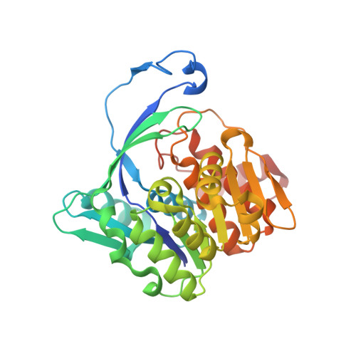
Structure of Burkholderia thailandensis nucleoside kinase (BthNK) in complex with AMP-Mg-AMP


Experimental Data Snapshot

  • Method: X-RAY DIFFRACTION
  • Resolution: 2.00 Å
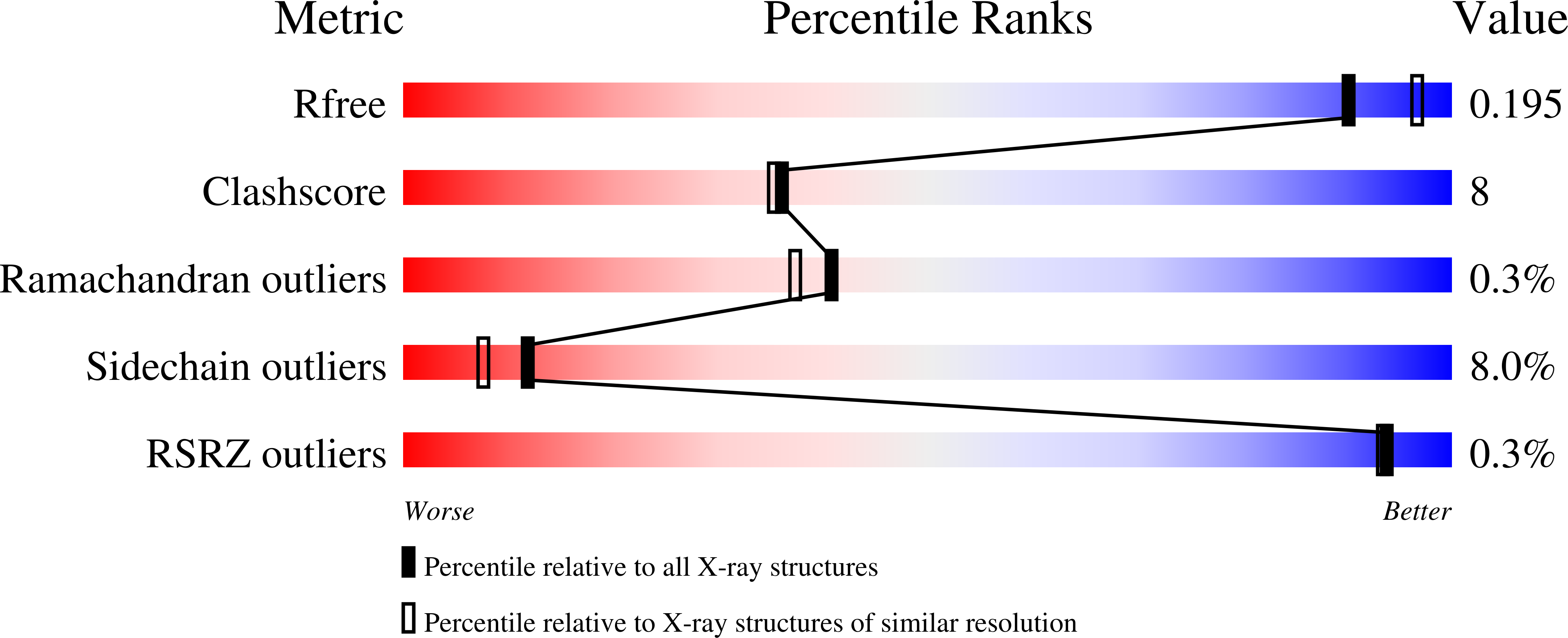
  • R-Value Free: 0.204 
  • R-Value Work: 0.167 
  • R-Value Observed: 0.169 

Starting Model: experimental
View more details

wwPDB Validation   3D Report Full Report


Ligand Structure Quality Assessment 


This is version 1.2 of the entry. See complete history


Literature

Structures of Burkholderia thailandensis nucleoside kinase: implications for the catalytic mechanism and nucleoside selectivity

Yasutake, Y.Ota, H.Hino, E.Sakasegawa, S.Tamura, T.

(2011) Acta Crystallogr D Biol Crystallogr 67: 945-956

  • DOI: https://doi.org/10.1107/S0907444911038777
  • Primary Citation of Related Structures:  
    3B1N, 3B1O, 3B1P, 3B1Q, 3B1R

  • PubMed Abstract: 

    The nucleoside kinase (NK) from the mesophilic Gram-negative bacterium Burkholderia thailandensis (BthNK) is a member of the phosphofructokinase B (Pfk-B) family and catalyzes the Mg(2+)- and ATP-dependent phosphorylation of a broad range of nucleosides such as inosine (INO), adenosine (ADO) and mizoribine (MZR). BthNK is currently used in clinical practice to measure serum MZR levels. Here, crystal structures of BthNK in a ligand-free form and in complexes with INO, INO-ADP, MZR-ADP and AMP-Mg(2+)-AMP are described. The typical homodimeric architecture of Pfk-B enzymes was detected in three distinct conformational states: an asymmetric dimer with one subunit in an open conformation and the other in a closed conformation (the ligand-free form), a closed conformation (the binary complex with INO) and a fully closed conformation (the other ternary and quaternary complexes). The previously unreported fully closed structures suggest the possibility that Mg(2+) might directly interact with the β- and γ-phosphates of ATP to maintain neutralization of the negative charge throughout the reaction. The nucleoside-complex structures also showed that the base moiety of the bound nucleoside is partly exposed to the solvent, thereby enabling the recognition of a wide range of nucleoside bases. Gly170 is responsible for the solvent accessibility of the base moiety and is assumed to be a key residue for the broad nucleoside recognition of BthNK. Remarkably, the G170Q mutation increases the specificity of BthNK for ADO. These findings provide insight into the conformational dynamics, catalytic mechanism and nucleoside selectivity of BthNK and related enzymes.


  • Organizational Affiliation

    Bioproduction Research Institute, National Institute of Advanced Industrial Science and Technology (AIST), 2-17-2-1 Tsukisamu-Higashi, Sapporo 062-8517, Japan.


Macromolecules
Find similar proteins by:  (by identity cutoff)  |  3D Structure
Entity ID: 1
MoleculeChains Sequence LengthOrganismDetailsImage
Ribokinase, putative
A, B, C, D, E
A, B, C, D, E, F
320Burkholderia thailandensis E264Mutation(s): 0 
Gene Names: BTH_I1158
EC: 2.7.1.143
UniProt
Find proteins for Q2SZE4 (Burkholderia thailandensis (strain ATCC 700388 / DSM 13276 / CCUG 48851 / CIP 106301 / E264))
Explore Q2SZE4 
Go to UniProtKB:  Q2SZE4
Entity Groups  
Sequence Clusters30% Identity50% Identity70% Identity90% Identity95% Identity100% Identity
UniProt GroupQ2SZE4
Sequence Annotations
Expand
  • Reference Sequence
Small Molecules
Ligands 2 Unique
IDChains Name / Formula / InChI Key2D Diagram3D Interactions
AMP
Query on AMP

Download Ideal Coordinates CCD File 
G [auth A]
H [auth A]
J [auth B]
K [auth B]
M [auth C]
G [auth A],
H [auth A],
J [auth B],
K [auth B],
M [auth C],
N [auth C],
P [auth D],
Q [auth D],
T [auth E],
U [auth E],
W [auth F],
X [auth F]
ADENOSINE MONOPHOSPHATE
C10 H14 N5 O7 P
UDMBCSSLTHHNCD-KQYNXXCUSA-N
MG
Query on MG

Download Ideal Coordinates CCD File 
I [auth A]
L [auth B]
O [auth C]
R [auth D]
S [auth D]
I [auth A],
L [auth B],
O [auth C],
R [auth D],
S [auth D],
V [auth E],
Y [auth F]
MAGNESIUM ION
Mg
JLVVSXFLKOJNIY-UHFFFAOYSA-N
Experimental Data & Validation

Experimental Data

  • Method: X-RAY DIFFRACTION
  • Resolution: 2.00 Å
  • R-Value Free: 0.204 
  • R-Value Work: 0.167 
  • R-Value Observed: 0.169 
  • Space Group: P 31
Unit Cell:
Length ( Å )Angle ( ˚ )
a = 125.222α = 90
b = 125.222β = 90
c = 115.002γ = 120
Software Package:
Software NamePurpose
ADSCdata collection
PHASERphasing
REFMACrefinement
HKL-2000data reduction
HKL-2000data scaling

Structure Validation

View Full Validation Report


Ligand Structure Quality Assessment 


Entry History 

Deposition Data

Revision History  (Full details and data files)

  • Version 1.0: 2011-11-02
    Type: Initial release
  • Version 1.1: 2012-04-18
    Changes: Database references
  • Version 1.2: 2023-11-01
    Changes: Data collection, Database references, Derived calculations, Refinement description