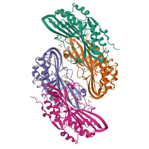
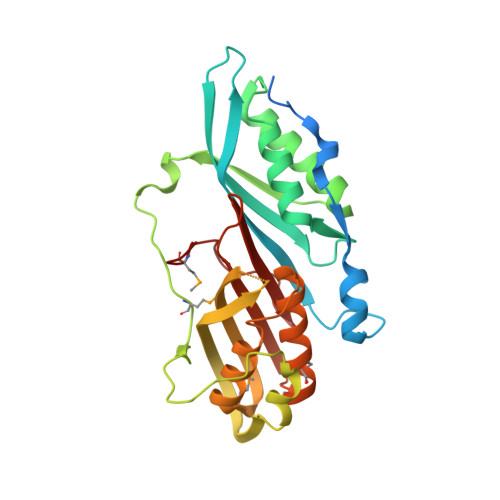
High-resolution structure of the nitrile reductase QueF combined with molecular simulations provide insight into enzyme mechanism.
Kim, Y., Zhou, M., Moy, S., Morales, J., Cunningham, M.A., Joachimiak, A.(2010) J Mol Biology 404: 127-137
- PubMed: 20875425
- DOI: https://doi.org/10.1016/j.jmb.2010.09.042
- Primary Citation of Related Structures:
3BP1 - PubMed Abstract:
Here, we report the 1.53-Å crystal structure of the enzyme 7-cyano-7-deazaguanine reductase (QueF) from Vibrio cholerae, which is responsible for the complete reduction of a nitrile (CN) bond to a primary amine (H(2)C-NH(2)). At present, this is the only example of a biological pathway that includes reduction of a nitrile bond, establishing QueF as particularly noteworthy. The structure of the QueF monomer resembles two connected ferrodoxin-like domains that assemble into dimers. Ligands identified in the crystal structure suggest the likely binding conformation of the native substrates NADPH and 7-cyano-7-deazaguanine. We also report on a series of numerical simulations that have shed light on the mechanism by which this enzyme affects the transfer of four protons (and electrons) to the 7-cyano-7-deazaguanine substrate. In particular, the simulations suggest that the initial step of the catalytic process is the formation of a covalent adduct with the residue Cys194, in agreement with previous studies. The crystal structure also suggests that two conserved residues (His233 and Asp102) play an important role in the delivery of a fourth proton to the substrate.
Organizational Affiliation:
The Midwest Center for Structural Genomics and Structural Biology Center, Biosciences, Argonne National Laboratory, 9700 South Cass Avenue, Argonne, IL 60439, USA.


