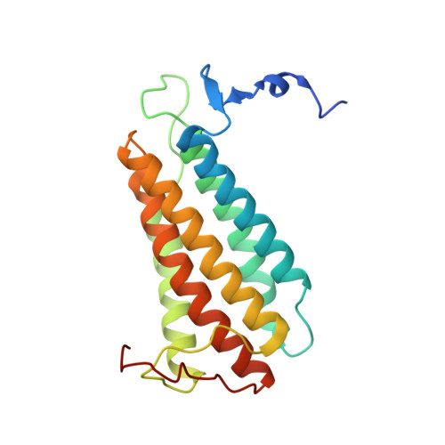
Structural characterization of a human-type corrinoid adenosyltransferase confirms that coenzyme B12 is synthesized through a four-coordinate intermediate.
St Maurice, M., Mera, P., Park, K., Brunold, T.C., Escalante-Semerena, J.C., Rayment, I.(2008) Biochemistry 47: 5755-5766
- PubMed: 18452306
- DOI: https://doi.org/10.1021/bi800132d
- Primary Citation of Related Structures:
3CI1, 3CI3, 3CI4 - PubMed Abstract:
ATP:cob(I)alamin adenosyltransferases (ACAs) catalyze the transfer of the 5'-deoxyadenosyl moiety from ATP to the upper axial ligand position of cobalamin in the synthesis of coenzyme B 12. For the ACA-catalyzed reaction to proceed, cob(II)alamin must be reduced to cob(I)alamin in the enzyme active site. This reduction is facilitated through the generation of a four-coordinate cob(II)alamin intermediate on the enzyme. We have determined the high-resolution crystal structure of a human-type ACA from Lactobacillus reuteri with a four-coordinate cob(II)alamin bound in the enzyme active site and with the product, adenosylcobalamin, partially occupied in the active site. The assembled structures represent snapshots of the steps in the ACA-catalyzed formation of the cobalt-carbon bond of coenzyme B 12. The structures define the corrinoid binding site and provide visual evidence for a base-off, four-coordinate cob(II)alamin intermediate. The complete structural description of ACA-mediated catalysis reveals the molecular features of four-coordinate cob(II)alamin stabilization and provides additional insights into the molecular basis for dysfunction in human patients suffering from methylmalonic aciduria.
Organizational Affiliation:
Departments of Biochemistry, University of Wisconsin, Madison, Wisconsin 53706, USA.


