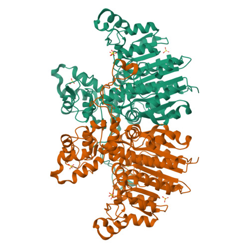
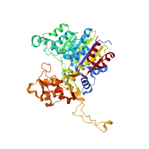
3E2D

The 1.4 A crystal structure of the large and cold-active Vibrio sp. alkaline phosphatase


Experimental Data Snapshot

  • Method: X-RAY DIFFRACTION
  • Resolution: 1.40 Å
  • R-Value Free: 0.166 
  • R-Value Work: 0.155 
  • R-Value Observed: 0.156 

wwPDB Validation   3D Report Full Report


This is version 1.3 of the entry. See complete history


Literature

The 1.4 A crystal structure of the large and cold-active Vibrio sp. alkaline phosphatase.

Helland, R.Larsen, R.L.Asgeirsson, B.

(2009) Biochim Biophys Acta 1794: 297-308

  • DOI: https://doi.org/10.1016/j.bbapap.2008.09.020
  • Primary Citation of Related Structures:  
    3E2D

  • PubMed Abstract: 

    Alkaline phosphatase (AP) from the cold-adapted Vibrio strain G15-21 is among the AP variants with the highest known k(cat) value. Here the structure of the enzyme at 1.4 A resolution is reported and compared to APs from E. coli, human placenta, shrimp and the Antarctic bacterium strain TAB5. The Vibrio AP is a dimer although its monomers are without the long N-terminal helix that embraces the other subunit in many other APs. The long insertion loop, previously noted as a special feature of the Vibrio AP, serves a similar function. The surface does not have the high negative charge density as observed in shrimp AP, but a positively charged patch is observed around the active site that may be favourable for substrate binding. The dimer interface has a similar number of non-covalent interactions as other APs and the "crown"-domain is the largest observed in known APs. Part of it slopes over the catalytic site suggesting that the substrates may be small molecules. The catalytic serines are refined with multiple conformations in both monomers. One of the ligands to the catalytic zinc ion in binding site M1 is directly connected to the crown-domain and is closest to the dimer interface. Subtle movements in metal ligands may help in the release of the product and/or facilitate prior dephosphorylation of the covalent intermediate. Intersubunit interactions may be a major factor for promoting active site geometries that lead to the high catalytic activity of Vibrio AP at low temperatures.


  • Organizational Affiliation

    The Norwegian Structural Biology Centre, Department of Chemistry, University of Tromsø, N-9037 Tromsø, Norway.


Macromolecules
Find similar proteins by:  (by identity cutoff)  |  3D Structure
Entity ID: 1
MoleculeChains Sequence LengthOrganismDetailsImage
Alkaline phosphatase
A, B
502Vibrio sp. G15-21Mutation(s): 0 
EC: 3.1.3.1
UniProt
Find proteins for Q93P54 (Vibrio sp. G15-21)
Explore Q93P54 
Go to UniProtKB:  Q93P54
Entity Groups  
Sequence Clusters30% Identity50% Identity70% Identity90% Identity95% Identity100% Identity
UniProt GroupQ93P54
Sequence Annotations
Expand
  • Reference Sequence
Small Molecules
Ligands 4 Unique
IDChains Name / Formula / InChI Key2D Diagram3D Interactions
SO4
Query on SO4

Download Ideal Coordinates CCD File 
F [auth A]
G [auth A]
H [auth A]
I [auth A]
J [auth A]
F [auth A],
G [auth A],
H [auth A],
I [auth A],
J [auth A],
K [auth A],
R [auth B],
S [auth B],
T [auth B],
U [auth B],
V [auth B]
SULFATE ION
O4 S
QAOWNCQODCNURD-UHFFFAOYSA-L
ZN
Query on ZN

Download Ideal Coordinates CCD File 
C [auth A],
D [auth A],
O [auth B],
P [auth B]
ZINC ION
Zn
PTFCDOFLOPIGGS-UHFFFAOYSA-N
EDO
Query on EDO

Download Ideal Coordinates CCD File 
L [auth A],
M [auth A],
N [auth A]
1,2-ETHANEDIOL
C2 H6 O2
LYCAIKOWRPUZTN-UHFFFAOYSA-N
MG
Query on MG

Download Ideal Coordinates CCD File 
E [auth A],
Q [auth B]
MAGNESIUM ION
Mg
JLVVSXFLKOJNIY-UHFFFAOYSA-N
Experimental Data & Validation

Experimental Data

  • Method: X-RAY DIFFRACTION
  • Resolution: 1.40 Å
  • R-Value Free: 0.166 
  • R-Value Work: 0.155 
  • R-Value Observed: 0.156 
  • Space Group: P 21 21 2
Unit Cell:
Length ( Å )Angle ( ˚ )
a = 118.24α = 90
b = 165.98β = 90
c = 57.48γ = 90
Software Package:
Software NamePurpose
REFMACrefinement
MAR345data collection
XDSdata reduction
SCALAdata scaling
SHELXDphasing
SHARPphasing

Structure Validation

View Full Validation Report


Entry History 

Deposition Data

Revision History  (Full details and data files)

  • Version 1.0: 2009-06-16
    Type: Initial release
  • Version 1.1: 2011-07-13
    Changes: Version format compliance
  • Version 1.2: 2017-10-25
    Changes: Refinement description
  • Version 1.3: 2024-03-20
    Changes: Data collection, Database references, Derived calculations