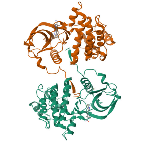
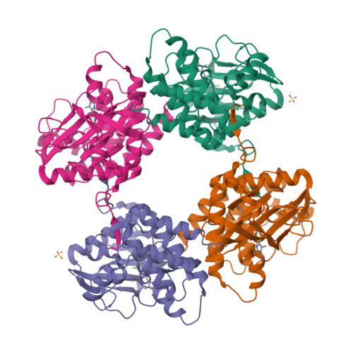
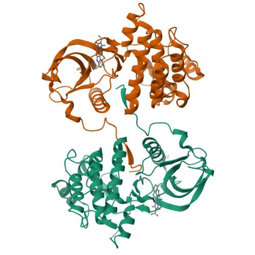
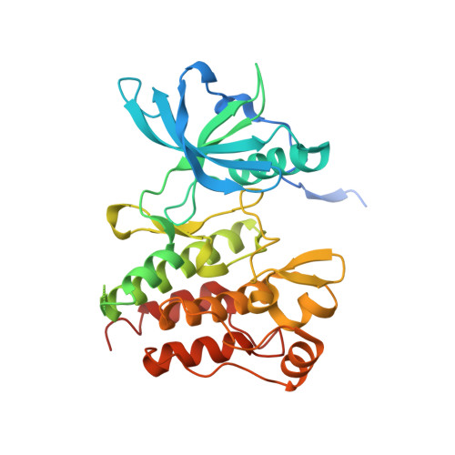
3G0F

KIT kinase domain mutant D816H in complex with sunitinib


Experimental Data Snapshot

  • Method: X-RAY DIFFRACTION
  • Resolution: 2.60 Å
  • R-Value Free: 0.255 
  • R-Value Work: 0.204 
  • R-Value Observed: 0.207 

Starting Model: experimental
View more details

wwPDB Validation   3D Report Full Report


Ligand Structure Quality Assessment 


This is version 1.5 of the entry. See complete history


Literature

KIT kinase mutants show unique mechanisms of drug resistance to imatinib and sunitinib in gastrointestinal stromal tumor patients.

Gajiwala, K.S.Wu, J.C.Christensen, J.Deshmukh, G.D.Diehl, W.DiNitto, J.P.English, J.M.Greig, M.J.He, Y.A.Jacques, S.L.Lunney, E.A.McTigue, M.Molina, D.Quenzer, T.Wells, P.A.Yu, X.Zhang, Y.Zou, A.Emmett, M.R.Marshall, A.G.Zhang, H.M.Demetri, G.D.

(2009) Proc Natl Acad Sci U S A 106: 1542-1547

  • DOI: https://doi.org/10.1073/pnas.0812413106
  • Primary Citation of Related Structures:  
    3G0E, 3G0F

  • PubMed Abstract: 

    Most gastrointestinal stromal tumors (GISTs) exhibit aberrant activation of the receptor tyrosine kinase (RTK) KIT. The efficacy of the inhibitors imatinib mesylate and sunitinib malate in GIST patients has been linked to their inhibition of these mutant KIT proteins. However, patients on imatinib can acquire secondary KIT mutations that render the protein insensitive to the inhibitor. Sunitinib has shown efficacy against certain imatinib-resistant mutants, although a subset that resides in the activation loop, including D816H/V, remains resistant. Biochemical and structural studies were undertaken to determine the molecular basis of sunitinib resistance. Our results show that sunitinib targets the autoinhibited conformation of WT KIT and that the D816H mutant undergoes a shift in conformational equilibrium toward the active state. These findings provide a structural and enzymologic explanation for the resistance profile observed with the KIT inhibitors. Prospectively, they have implications for understanding oncogenic kinase mutants and for circumventing drug resistance.


  • Organizational Affiliation

    Departments of Structural and Computational Biology, Cancer Biology, and Biochemical Pharmacology, Pfizer Global Research and Development, 10777 Science Center Drive, La Jolla, CA 92121, USA.


Macromolecules
Find similar proteins by:  (by identity cutoff)  |  3D Structure
Entity ID: 1
MoleculeChains Sequence LengthOrganismDetailsImage
Mast/stem cell growth factor receptor
A, B
336Homo sapiensMutation(s): 1 
Gene Names: KIT
EC: 2.7.10.1
UniProt & NIH Common Fund Data Resources
Find proteins for P10721 (Homo sapiens)
Explore P10721 
Go to UniProtKB:  P10721
PHAROS:  P10721
GTEx:  ENSG00000157404 
Entity Groups  
Sequence Clusters30% Identity50% Identity70% Identity90% Identity95% Identity100% Identity
UniProt GroupP10721
Sequence Annotations
Expand
  • Reference Sequence
Small Molecules
Ligands 2 Unique
IDChains Name / Formula / InChI Key2D Diagram3D Interactions
B49
Query on B49

Download Ideal Coordinates CCD File 
C [auth A],
E [auth B]
N-[2-(diethylamino)ethyl]-5-[(Z)-(5-fluoro-2-oxo-1,2-dihydro-3H-indol-3-ylidene)methyl]-2,4-dimethyl-1H-pyrrole-3-carbo xamide
C22 H27 F N4 O2
WINHZLLDWRZWRT-ATVHPVEESA-N
SO4
Query on SO4

Download Ideal Coordinates CCD File 
D [auth A]SULFATE ION
O4 S
QAOWNCQODCNURD-UHFFFAOYSA-L
Binding Affinity Annotations 
IDSourceBinding Affinity
B49 BindingDB:  3G0F Ki: min: 4, max: 410 (nM) from 5 assay(s)
Kd: min: 0.21, max: 380 (nM) from 13 assay(s)
IC50: min: 1.1, max: 3021 (nM) from 11 assay(s)
PDBBind:  3G0F Kd: 22 (nM) from 1 assay(s)
Experimental Data & Validation

Experimental Data

  • Method: X-RAY DIFFRACTION
  • Resolution: 2.60 Å
  • R-Value Free: 0.255 
  • R-Value Work: 0.204 
  • R-Value Observed: 0.207 
  • Space Group: P 21 21 2
Unit Cell:
Length ( Å )Angle ( ˚ )
a = 79.462α = 90
b = 101.783β = 90
c = 105.505γ = 90
Software Package:
Software NamePurpose
ADSCdata collection
CNXrefinement
HKL-2000data reduction
SCALEPACKdata scaling
CNXphasing

Structure Validation

View Full Validation Report


Ligand Structure Quality Assessment 


Entry History 

Deposition Data

Revision History  (Full details and data files)

  • Version 1.0: 2009-02-24
    Type: Initial release
  • Version 1.1: 2011-07-13
    Changes: Version format compliance
  • Version 1.2: 2017-08-16
    Changes: Refinement description, Source and taxonomy
  • Version 1.3: 2021-10-20
    Changes: Database references, Derived calculations, Structure summary
  • Version 1.4: 2023-11-29
    Changes: Data collection, Database references
  • Version 1.5: 2024-04-03
    Changes: Refinement description