3G3E | pdb_00003g3e

Crystal structure of human D-amino acid oxidase in complex with hydroxyquinolin-2(1H)


Experimental Data Snapshot

  • Method: X-RAY DIFFRACTION
  • Resolution: 2.20 Å
  • R-Value Free: 
    0.294 (Depositor), 0.300 (DCC) 
  • R-Value Work: 
    0.239 (Depositor), 0.250 (DCC) 
  • R-Value Observed: 
    0.242 (Depositor) 

Starting Model: experimental
View more details

wwPDB Validation   3D Report Full Report


Ligand Structure Quality Assessment 

Created with Raphaël 2.3.0Worse 01 BetterLigand structure goodness of fit to experimental dataBest fitted FADClick on this verticalbar to view detailsBest fitted G3EClick on this verticalbar to view details

This is version 1.2 of the entry. See complete history


Literature

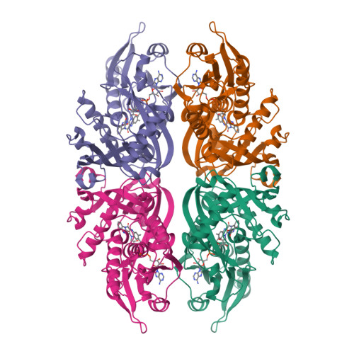
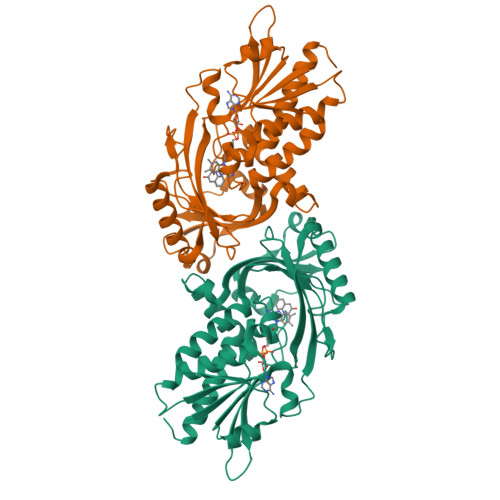
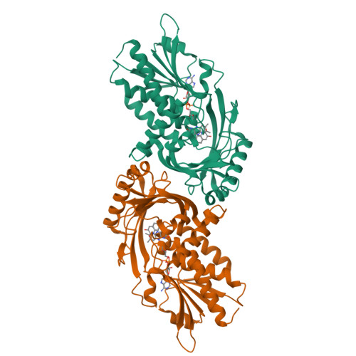
Discovery, SAR, and pharmacokinetics of a novel 3-Hydroxyquinolin-2(1H)-one series of potent D-amino acid oxidase (DAAO) inhibitors

Duplantier, A.J.Becker, S.L.Bohanon, M.J.Borzilleri, K.A.Chrunyk, B.A.Downs, J.T.Hu, L.Y.El-Kattan, A.James, L.C.Liu, S.Lu, J.Maklad, N.Mansour, M.N.Mente, S.Piotrowski, M.A.Sakya, S.M.Sheehan, S.Steyn, S.J.Strick, C.A.Williams, V.A.Zhang, L.

(2009) J Med Chem 52: 3576-3585

  • DOI: https://doi.org/10.1021/jm900128w
  • Primary Citation of Related Structures:  
    3G3E

  • PubMed Abstract: 

    3-Hydroxyquinolin-2(1H)-one (2) was discovered by high throughput screening in a functional assay to be a potent inhibitor of human DAAO, and its binding affinity was confirmed in a Biacore assay. Cocrystallization of 2 with the human DAAO enzyme defined the binding site and guided the design of new analogues. The SAR, pharmacokinetics, brain exposure, and effects on cerebellum D-serine are described. Subsequent evaluation against the rat DAAO enzyme revealed a divergent SAR versus the human enzyme and may explain the high exposures of drug necessary to achieve significant changes in rat or mouse cerebellum D-serine.


  • Organizational Affiliation

    Pfizer Global Research and Development, Groton Laboratories, Groton, CT 06340, USA. allen.j.duplantier@pfizer.com


Macromolecules
Find similar proteins by:  (by identity cutoff)  |  3D Structure
Entity ID: 1
MoleculeChains Sequence LengthOrganismDetailsImage
D-amino-acid oxidase
A, B, C, D
351Homo sapiensMutation(s): 0 
Gene Names: DAODAMOX
EC: 1.4.3.3
UniProt & NIH Common Fund Data Resources
Find proteins for P14920 (Homo sapiens)
Explore P14920 
Go to UniProtKB:  P14920
PHAROS:  P14920
GTEx:  ENSG00000110887 
Entity Groups  
Sequence Clusters30% Identity50% Identity70% Identity90% Identity95% Identity100% Identity
UniProt GroupP14920
Sequence Annotations
Expand
  • Reference Sequence
Small Molecules
Ligands 2 Unique
IDChains Name / Formula / InChI Key2D Diagram3D Interactions
FAD
Query on FAD

Download Ideal Coordinates CCD File 
E [auth A],
G [auth B],
I [auth C],
K [auth D]
FLAVIN-ADENINE DINUCLEOTIDE
C27 H33 N9 O15 P2
VWWQXMAJTJZDQX-UYBVJOGSSA-N
G3E
Query on G3E

Download Ideal Coordinates CCD File 
F [auth A],
H [auth B],
J [auth C],
L [auth D]
3-hydroxyquinolin-2(1H)-one
C9 H7 N O2
BERPCVULMUPOER-UHFFFAOYSA-N
Binding Affinity Annotations 
IDSourceBinding Affinity
G3E BindingDB:  3G3E Kd: 13 (nM) from 1 assay(s)
IC50: min: 4, max: 43 (nM) from 4 assay(s)
Experimental Data & Validation

Experimental Data

  • Method: X-RAY DIFFRACTION
  • Resolution: 2.20 Å
  • R-Value Free:  0.294 (Depositor), 0.300 (DCC) 
  • R-Value Work:  0.239 (Depositor), 0.250 (DCC) 
  • R-Value Observed: 0.242 (Depositor) 
Space Group: P 1
Unit Cell:
Length ( Å )Angle ( ˚ )
a = 51.063α = 102.62
b = 82.584β = 105.43
c = 96.974γ = 110.79
Software Package:
Software NamePurpose
REFMACrefinement

Structure Validation

View Full Validation Report


Ligand Structure Quality Assessment 

Created with Raphaël 2.3.0Worse 01 BetterLigand structure goodness of fit to experimental dataBest fitted FADClick on this verticalbar to view detailsBest fitted G3EClick on this verticalbar to view details

Entry History 

Deposition Data

Revision History  (Full details and data files)

  • Version 1.0: 2009-06-23
    Type: Initial release
  • Version 1.1: 2011-07-13
    Changes: Advisory, Version format compliance
  • Version 1.2: 2023-09-06
    Changes: Data collection, Database references, Derived calculations, Refinement description