3G51 | pdb_00003g51

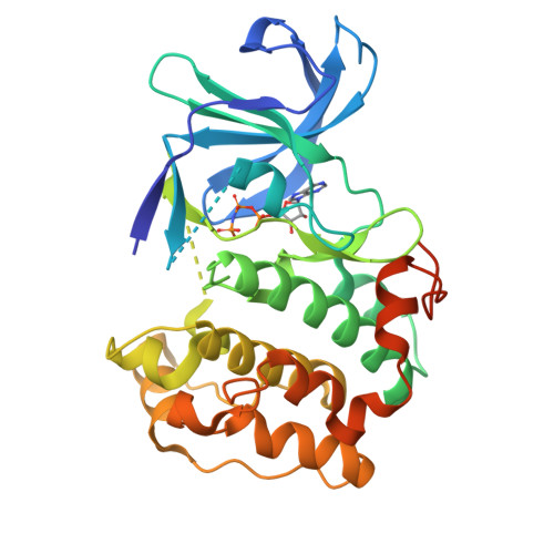
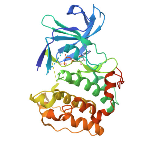
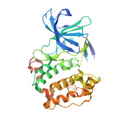
Structural diversity of the active conformation of the N-terminal kinase domain of p90 ribosomal S6 kinase 2


Experimental Data Snapshot

  • Method: X-RAY DIFFRACTION
  • Resolution: 1.80 Å
  • R-Value Free: 
    0.248 (Depositor) 
  • R-Value Work: 
    0.204 (Depositor), 0.210 (DCC) 
  • R-Value Observed: 
    0.216 (Depositor) 

Starting Model: experimental
View more details

wwPDB Validation   3D Report Full Report


Ligand Structure Quality Assessment 

Created with Raphaël 2.3.0Worse 01 BetterLigand structure goodness of fit to experimental dataBest fitted ANPClick on this verticalbar to view details

This is version 1.2 of the entry. See complete history


Literature

Structural diversity of the active N-terminal kinase domain of p90 ribosomal S6 kinase 2

Malakhova, M.Kurinov, I.Liu, K.Zheng, D.D'Angelo, I.Shim, J.H.Steinman, V.Bode, A.M.Dong, Z.

(2009) PLoS One 4: e8044-e8044

  • DOI: https://doi.org/10.1371/journal.pone.0008044
  • Primary Citation of Related Structures:  
    3G51

  • PubMed Abstract: 

    The p90 ribosomal protein kinase 2 (RSK2) is a highly expressed Ser/Thr kinase activated by growth factors and is involved in cancer cell proliferation and tumor promoter-induced cell transformation. RSK2 possesses two non-identical kinase domains, and the structure of its N-terminal domain (NTD), which is responsible for phosphorylation of a variety of substrates, is unknown. The crystal structure of the NTD RSK2 was determined at 1.8 A resolution in complex with AMP-PNP. The N-terminal kinase domain adopted a unique active conformation showing a significant structural diversity of the kinase domain compared to other kinases. The NTD RSK2 possesses a three-stranded betaB-sheet inserted in the N-terminal lobe, resulting in displacement of the alphaC-helix and disruption of the Lys-Glu interaction, classifying the kinase conformation as inactive. The purified protein was phosphorylated at Ser227 in the T-activation loop and exhibited in vitro kinase activity. A key characteristic is the appearance of a new contact between Lys216 (betaB-sheet) and the beta-phosphate of AMP-PNP. Mutation of this lysine to alanine impaired both NTDs in vitro and full length RSK2 ex vivo activity, emphasizing the importance of this interaction. Even though the N-terminal lobe undergoes structural re-arrangement, it possesses an intact hydrophobic groove formed between the alphaC-helix, the beta4-strand, and the betaB-sheet junction, which is occupied by the N-terminal tail. The presence of a unique betaB-sheet insert in the N-lobe suggests a different type of activation mechanism for RSK2.


  • Organizational Affiliation

    Department of Cellular and Molecular Biology, The Hormel Institute, University of Minnesota, Austin, Minnesota, United States of America.


Macromolecules
Find similar proteins by:  (by identity cutoff)  |  3D Structure
Entity ID: 1
MoleculeChains Sequence LengthOrganismDetailsImage
Ribosomal protein S6 kinase alpha-3325Mus musculusMutation(s): 0 
Gene Names: Mus musculus ribosomal protein S6 kinase 2Rps6ka-rs1Rps6ka3Rsk2
EC: 2.7.11.1
UniProt
Find proteins for P18654 (Mus musculus)
Explore P18654 
Go to UniProtKB:  P18654
Entity Groups  
Sequence Clusters30% Identity50% Identity70% Identity90% Identity95% Identity100% Identity
UniProt GroupP18654
Sequence Annotations
Expand
  • Reference Sequence
Small Molecules
Ligands 1 Unique
IDChains Name / Formula / InChI Key2D Diagram3D Interactions
ANP
Query on ANP

Download Ideal Coordinates CCD File 
B [auth A]PHOSPHOAMINOPHOSPHONIC ACID-ADENYLATE ESTER
C10 H17 N6 O12 P3
PVKSNHVPLWYQGJ-KQYNXXCUSA-N
Experimental Data & Validation

Experimental Data

  • Method: X-RAY DIFFRACTION
  • Resolution: 1.80 Å
  • R-Value Free:  0.248 (Depositor) 
  • R-Value Work:  0.204 (Depositor), 0.210 (DCC) 
  • R-Value Observed: 0.216 (Depositor) 
Space Group: P 21 21 21
Unit Cell:
Length ( Å )Angle ( ˚ )
a = 51.167α = 90
b = 51.381β = 90
c = 140.873γ = 90
Software Package:
Software NamePurpose
ADSCdata collection
PHENIXmodel building
PHENIXrefinement
HKL-2000data reduction
HKL-2000data scaling
PHENIXphasing

Structure Validation

View Full Validation Report


Ligand Structure Quality Assessment 

Created with Raphaël 2.3.0Worse 01 BetterLigand structure goodness of fit to experimental dataBest fitted ANPClick on this verticalbar to view details

Entry History 

Deposition Data

  • Released Date: 2009-12-15 
  • Deposition Author(s): Kurinov, I.

Revision History  (Full details and data files)

  • Version 1.0: 2009-12-15
    Type: Initial release
  • Version 1.1: 2011-07-13
    Changes: Version format compliance
  • Version 1.2: 2023-09-06
    Changes: Data collection, Database references, Derived calculations, Refinement description