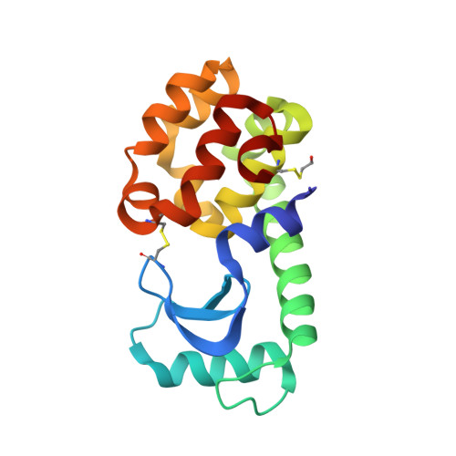
3GUP

T4 lysozyme M102E/L99A mutant with buried charge in apolar cavity--pyridine binding


Experimental Data Snapshot

  • Method: X-RAY DIFFRACTION
  • Resolution: 1.50 Å
  • R-Value Free: 0.253 
  • R-Value Work: 0.211 
  • R-Value Observed: 0.213 

Starting Model: experimental
View more details

wwPDB Validation   3D Report Full Report


This is version 1.5 of the entry. See complete history


Literature

Use of stabilizing mutations to engineer a charged group within a ligand-binding hydrophobic cavity in T4 lysozyme.

Liu, L.Baase, W.A.Michael, M.M.Matthews, B.W.

(2009) Biochemistry 48: 8842-8851

  • DOI: https://doi.org/10.1021/bi900685j
  • Primary Citation of Related Structures:  
    3GUI, 3GUJ, 3GUK, 3GUL, 3GUM, 3GUN, 3GUO, 3GUP

  • PubMed Abstract: 

    Both large-to-small and nonpolar-to-polar mutations in the hydrophobic core of T4 lysozyme cause significant loss in stability. By including supplementary stabilizing mutations we constructed a variant that combines the cavity-creating substitution Leu99 --> Ala with the buried charge mutant Met102 --> Glu. Crystal structure determination confirmed that this variant has a large cavity with the side chain of Glu102 located within the cavity wall. The cavity includes a large disk-shaped region plus a bulge. The disk-like region is essentially nonpolar, similar to L99A, while the Glu102 substituent is located in the vicinity of the bulge. Three ordered water molecules bind within this part of the cavity and appear to stabilize the conformation of Glu102. Glu102 has an estimated pKa of about 5.5-6.5, suggesting that it is at least partially charged in the crystal structure. The polar ligands pyridine, phenol and aniline bind within the cavity, and crystal structures of the complexes show one or two water molecules to be retained. Nonpolar ligands of appropriate shape can also bind in the cavity and in some cases exclude all three water molecules. This disrupts the hydrogen-bond network and causes the Glu102 side chain to move away from the ligand by up to 0.8 A where it remains buried in a completely nonpolar environment. Isothermal titration calorimetry revealed that the binding of these compounds stabilizes the protein by 4-6 kcal/mol. For both polar and nonpolar ligands the binding is enthalpically driven. Large negative changes in entropy adversely balance the binding of the polar ligands, whereas entropy has little effect on the nonpolar ligand binding.


  • Organizational Affiliation

    Institute of Molecular Biology and Department of Physics, 1229 University of Oregon, Eugene, Oregon 97403-1229, USA.


Macromolecules
Find similar proteins by:  (by identity cutoff)  |  3D Structure
Entity ID: 1
MoleculeChains Sequence LengthOrganismDetailsImage
Lysozyme
A, B
164Tequatrovirus T4Mutation(s): 8 
Gene Names: E
EC: 3.2.1.17
UniProt
Find proteins for P00720 (Enterobacteria phage T4)
Explore P00720 
Go to UniProtKB:  P00720
Entity Groups  
Sequence Clusters30% Identity50% Identity70% Identity90% Identity95% Identity100% Identity
UniProt GroupP00720
Sequence Annotations
Expand
  • Reference Sequence
Small Molecules
Ligands 4 Unique
IDChains Name / Formula / InChI Key2D Diagram3D Interactions
0PY
Query on 0PY

Download Ideal Coordinates CCD File 
C [auth A],
H [auth A],
I [auth B]
pyridine
C5 H5 N
JUJWROOIHBZHMG-UHFFFAOYSA-N
CO3
Query on CO3

Download Ideal Coordinates CCD File 
D [auth A],
J [auth B]
CARBONATE ION
C O3
BVKZGUZCCUSVTD-UHFFFAOYSA-L
CL
Query on CL

Download Ideal Coordinates CCD File 
F [auth A],
G [auth A],
K [auth B],
L [auth B]
CHLORIDE ION
Cl
VEXZGXHMUGYJMC-UHFFFAOYSA-M
MG
Query on MG

Download Ideal Coordinates CCD File 
E [auth A]MAGNESIUM ION
Mg
JLVVSXFLKOJNIY-UHFFFAOYSA-N
Modified Residues  1 Unique
IDChains TypeFormula2D DiagramParent
CME
Query on CME
A, B
L-PEPTIDE LINKINGC5 H11 N O3 S2CYS
Experimental Data & Validation

Experimental Data

  • Method: X-RAY DIFFRACTION
  • Resolution: 1.50 Å
  • R-Value Free: 0.253 
  • R-Value Work: 0.211 
  • R-Value Observed: 0.213 
  • Space Group: P 43
Unit Cell:
Length ( Å )Angle ( ˚ )
a = 48.957α = 90
b = 48.957β = 90
c = 129.315γ = 90
Software Package:
Software NamePurpose
Blu-Icedata collection
AMoREphasing
REFMACrefinement
HKL-2000data reduction
HKL-2000data scaling

Structure Validation

View Full Validation Report


Entry History 

Deposition Data

Revision History  (Full details and data files)

  • Version 1.0: 2009-08-25
    Type: Initial release
  • Version 1.1: 2011-07-13
    Changes: Advisory, Refinement description, Version format compliance
  • Version 1.2: 2017-11-01
    Changes: Refinement description
  • Version 1.3: 2021-10-13
    Changes: Database references, Derived calculations, Structure summary
  • Version 1.4: 2023-09-06
    Changes: Data collection, Refinement description
  • Version 1.5: 2024-11-20
    Changes: Structure summary