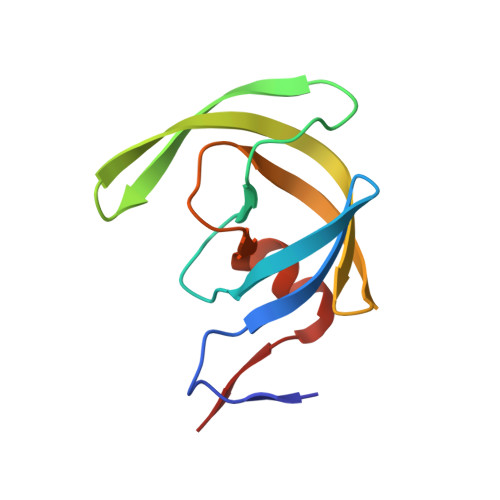
3H5B | pdb_00003h5b

Crystal structure of wild type HIV-1 protease with novel P1'-ligand GRL-02031


Experimental Data Snapshot

  • Method: X-RAY DIFFRACTION
  • Resolution: 1.29 Å
  • R-Value Free: 
    0.181 (Depositor), 0.180 (DCC) 
  • R-Value Work: 
    0.142 (Depositor), 0.150 (DCC) 
  • R-Value Observed: 
    0.144 (Depositor) 

Starting Model: experimental
View more details

wwPDB Validation   3D Report Full Report


Ligand Structure Quality Assessment 

Created with Raphaël 2.3.0Worse 01 BetterLigand structure goodness of fit to experimental dataBest fitted 031Click on this verticalbar to view details

This is version 1.4 of the entry. See complete history


Literature

Design of HIV-1 protease inhibitors with pyrrolidinones and oxazolidinones as novel P1'-ligands to enhance backbone-binding interactions with protease: synthesis, biological evaluation, and protein-ligand X-ray studies.

Ghosh, A.K.Leshchenko-Yashchuk, S.Anderson, D.D.Baldridge, A.Noetzel, M.Miller, H.B.Tie, Y.Wang, Y.F.Koh, Y.Weber, I.T.Mitsuya, H.

(2009) J Med Chem 52: 3902-3914

  • DOI: https://doi.org/10.1021/jm900303m
  • Primary Citation of Related Structures:  
    3H5B

  • PubMed Abstract: 

    Structure-based design, synthesis, and biological evaluation of a series of novel HIV-1 protease inhibitors are described. In an effort to enhance interactions with protease backbone atoms, we have incorporated stereochemically defined methyl-2-pyrrolidinone and methyl oxazolidinone as the P1'-ligands. These ligands are designed to interact with Gly-27' carbonyl and Arg-8 side chain in the S1'-subsite of the HIV protease. We have investigated the potential of these ligands in combination with our previously developed bis-tetrahydrofuran (bis-THF) and cyclopentanyltetrahydrofuran (Cp-THF) as the P2-ligands. Inhibitor 19b with a (R)-aminomethyl-2-pyrrolidinone and a Cp-THF was shown to be the most potent compound. This inhibitor maintained near full potency against multi-PI-resistant clinical HIV-1 variants. A high resolution protein-ligand X-ray crystal structure of 19b-bound HIV-1 protease revealed that the P1'-pyrrolidinone heterocycle and the P2-Cp-ligand are involved in several critical interactions with the backbone atoms in the S1' and S2 subsites of HIV-1 protease.


  • Organizational Affiliation

    Department of Chemistry, Purdue University, West Lafayette, Indiana 47907, USA. akghosh@purdue.edu


Macromolecules
Find similar proteins by:  (by identity cutoff)  |  3D Structure
Entity ID: 1
MoleculeChains Sequence LengthOrganismDetailsImage
HIV-1 protease
A, B
99Human immunodeficiency virus type 1 (BRU ISOLATE)Mutation(s): 5 
Gene Names: gag-pol
EC: 3.4.23.16
UniProt
Find proteins for P03367 (Human immunodeficiency virus type 1 group M subtype B (isolate BRU/LAI))
Explore P03367 
Go to UniProtKB:  P03367
Entity Groups  
Sequence Clusters30% Identity50% Identity70% Identity90% Identity95% Identity100% Identity
UniProt GroupP03367
Sequence Annotations
Expand
  • Reference Sequence
Small Molecules
Ligands 4 Unique
IDChains Name / Formula / InChI Key2D Diagram3D Interactions
031
Query on 031

Download Ideal Coordinates CCD File 
E [auth B](3aS,5R,6aR)-hexahydro-2H-cyclopenta[b]furan-5-yl [(1S,2R)-1-benzyl-2-hydroxy-3-([(4-methoxyphenyl)sulfonyl]{[(2R)-5-oxopyrrolidin-2-yl]methyl}amino)propyl]carbamate
C30 H39 N3 O8 S
RPIALZPTIFOQGC-CXLNPQPMSA-N
GOL
Query on GOL

Download Ideal Coordinates CCD File 
G [auth B]GLYCEROL
C3 H8 O3
PEDCQBHIVMGVHV-UHFFFAOYSA-N
CL
Query on CL

Download Ideal Coordinates CCD File 
D [auth A],
F [auth B]
CHLORIDE ION
Cl
VEXZGXHMUGYJMC-UHFFFAOYSA-M
NA
Query on NA

Download Ideal Coordinates CCD File 
C [auth A]SODIUM ION
Na
FKNQFGJONOIPTF-UHFFFAOYSA-N
Binding Affinity Annotations 
IDSourceBinding Affinity
031 BindingDB:  3H5B Ki: 0.1 (nM) from 1 assay(s)
Experimental Data & Validation

Experimental Data

  • Method: X-RAY DIFFRACTION
  • Resolution: 1.29 Å
  • R-Value Free:  0.181 (Depositor), 0.180 (DCC) 
  • R-Value Work:  0.142 (Depositor), 0.150 (DCC) 
  • R-Value Observed: 0.144 (Depositor) 
Space Group: P 21 21 2
Unit Cell:
Length ( Å )Angle ( ˚ )
a = 58.111α = 90
b = 86.416β = 90
c = 45.97γ = 90
Software Package:
Software NamePurpose
MAR345data collection
AMoREphasing
SHELXL-97refinement
HKL-2000data reduction
HKL-2000data scaling

Structure Validation

View Full Validation Report


Ligand Structure Quality Assessment 

Created with Raphaël 2.3.0Worse 01 BetterLigand structure goodness of fit to experimental dataBest fitted 031Click on this verticalbar to view details

Entry History 

Deposition Data

Revision History  (Full details and data files)

  • Version 1.0: 2009-06-16
    Type: Initial release
  • Version 1.1: 2011-07-13
    Changes: Non-polymer description, Version format compliance
  • Version 1.2: 2017-11-01
    Changes: Refinement description
  • Version 1.3: 2021-10-13
    Changes: Database references, Derived calculations
  • Version 1.4: 2023-09-06
    Changes: Data collection, Refinement description