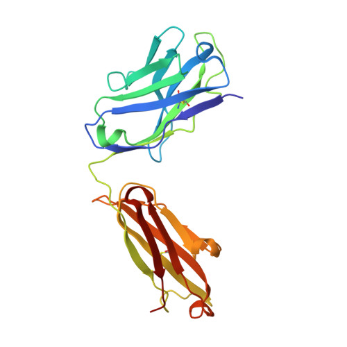
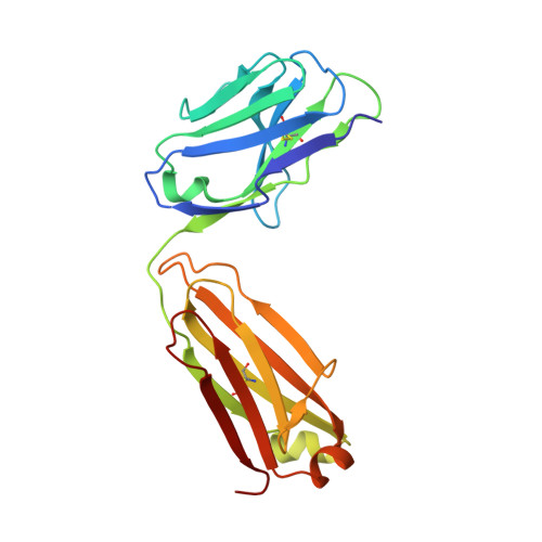
3I9G

Crystal structure of the LT1009 (SONEPCIZUMAB) antibody Fab fragment in complex with sphingosine-1-phosphate


Experimental Data Snapshot

  • Method: X-RAY DIFFRACTION
  • Resolution: 1.90 Å
  • R-Value Free: 0.219 
  • R-Value Work: 0.190 
  • R-Value Observed: 0.192 

wwPDB Validation   3D Report Full Report


Ligand Structure Quality Assessment 


This is version 1.2 of the entry. See complete history


Literature

The crystal structure of sphingosine-1-phosphate in complex with a Fab fragment reveals metal bridging of an antibody and its antigen.

Wojciak, J.M.Zhu, N.Schuerenberg, K.T.Moreno, K.Shestowsky, W.S.Hiraiwa, M.Sabbadini, R.Huxford, T.

(2009) Proc Natl Acad Sci U S A 106: 17717-17722

  • DOI: https://doi.org/10.1073/pnas.0906153106
  • Primary Citation of Related Structures:  
    3I9G

  • PubMed Abstract: 

    The pleiotropic signaling lipid sphingosine-1-phosphate (S1P) plays significant roles in angiogenesis, heart disease, and cancer. LT1009 (also known as sonepcizumab) is a humanized monoclonal antibody that binds S1P with high affinity and specificity. Because the antibody is currently in clinical trials, it is important to confirm by structural and biochemical analyses that it binds its target in a predictable manner. Therefore, we determined the structure of a complex between the LT1009 antibody Fab fragment and S1P refined to 1.90 A resolution. The antibody employs unique and diverse strategies to recognize its antigen. Two metal ions bridge complementarity determining regions from the antibody light chain and S1P. The coordination geometry, inductively coupled plasma spectroscopy, surface plasmon resonance spectroscopy, and biochemical assays suggest that these are Ca(2+). The amino alcohol head group of the sphingosine backbone is recognized through hydrogen bonding interactions from 1 aa side chain and polypeptide backbone atoms of the antibody light and heavy chains. The S1P hydrophobic tail is almost completely enclosed within a hydrophobic channel formed primarily by the heavy chain. Both treatment of the complex with metal chelators and mutation of amino acids in the light chain that coordinate the metal atoms or directly contact the polar head group abrogate binding, while mutations within the hydrophobic cavity also decrease S1P binding affinity. The structure suggests mechanistic details for recognition of a signaling lipid by a therapeutic antibody candidate. Moreover, this study provides direct structural evidence that antibodies are capable of using metals to bridge antigen:antibody complexes.


  • Organizational Affiliation

    Lpath Inc., 6335 Ferris Square, Suite A, San Diego, CA 92121, USA.


Macromolecules
Find similar proteins by:  (by identity cutoff)  |  3D Structure
Entity ID: 1
MoleculeChains Sequence LengthOrganismDetailsImage
Sonepcizumab antibody Fab fragment, heavy chainA [auth H]222Mus musculusMutation(s): 0 
Gene Names: IgG1K
Entity Groups  
Sequence Clusters30% Identity50% Identity70% Identity90% Identity95% Identity100% Identity
Sequence Annotations
Expand
  • Reference Sequence
Find similar proteins by:  (by identity cutoff)  |  3D Structure
Entity ID: 2
MoleculeChains Sequence LengthOrganismDetailsImage
Sonepcizumab antibody Fab fragment, light chainB [auth L]213Mus musculusMutation(s): 0 
Entity Groups  
Sequence Clusters30% Identity50% Identity70% Identity90% Identity95% Identity100% Identity
Sequence Annotations
Expand
  • Reference Sequence
Small Molecules
Ligands 3 Unique
IDChains Name / Formula / InChI Key2D Diagram3D Interactions
S1P
Query on S1P

Download Ideal Coordinates CCD File 
J [auth L](2S,3R,4E)-2-amino-3-hydroxyoctadec-4-en-1-yl dihydrogen phosphate
C18 H38 N O5 P
DUYSYHSSBDVJSM-KRWOKUGFSA-N
CA
Query on CA

Download Ideal Coordinates CCD File 
C [auth L],
D [auth L]
CALCIUM ION
Ca
BHPQYMZQTOCNFJ-UHFFFAOYSA-N
MG
Query on MG

Download Ideal Coordinates CCD File 
E [auth L],
F [auth L],
G [auth L],
H [auth L],
I [auth L]
MAGNESIUM ION
Mg
JLVVSXFLKOJNIY-UHFFFAOYSA-N
Binding Affinity Annotations 
IDSourceBinding Affinity
S1P PDBBind:  3I9G Kd: 1.1 (nM) from 1 assay(s)
Experimental Data & Validation

Experimental Data

  • Method: X-RAY DIFFRACTION
  • Resolution: 1.90 Å
  • R-Value Free: 0.219 
  • R-Value Work: 0.190 
  • R-Value Observed: 0.192 
  • Space Group: P 21 21 21
Unit Cell:
Length ( Å )Angle ( ˚ )
a = 66.052α = 90
b = 70.889β = 90
c = 138.719γ = 90
Software Package:
Software NamePurpose
PHASERphasing
REFMACrefinement
PDB_EXTRACTdata extraction
HKL-2000data collection
HKL-2000data reduction
HKL-2000data scaling

Structure Validation

View Full Validation Report


Ligand Structure Quality Assessment 


Entry History 

Deposition Data

  • Released Date: 2009-09-29 
  • Deposition Author(s): Huxford, T.

Revision History  (Full details and data files)

  • Version 1.0: 2009-09-29
    Type: Initial release
  • Version 1.1: 2011-07-13
    Changes: Version format compliance
  • Version 1.2: 2024-10-16
    Changes: Data collection, Database references, Derived calculations, Structure summary