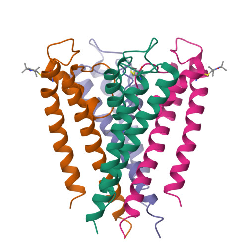
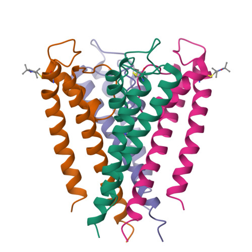
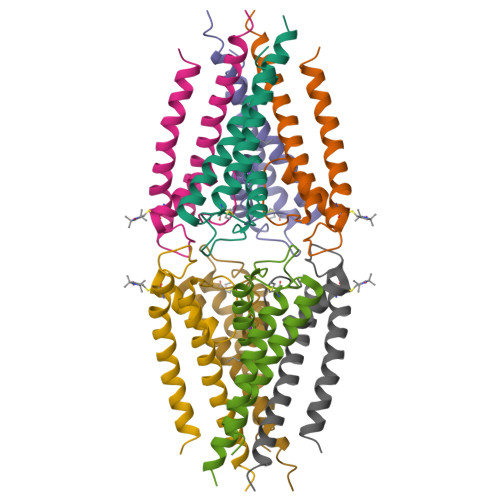
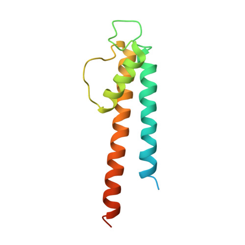
3IFX

Crystal structure of the Spin-labeled KcsA mutant V48R1


Experimental Data Snapshot

  • Method: X-RAY DIFFRACTION
  • Resolution: 3.56 Å
  • R-Value Free: 0.302 
  • R-Value Work: 0.271 
  • R-Value Observed: 0.273 

  • Method: EPR

Starting Model: experimental
View more details

wwPDB Validation   3D Report Full Report


Ligand Structure Quality Assessment 


This is version 1.3 of the entry. See complete history


Literature

Electron Spin-Echo Envelope Modulation (ESEEM) Reveals Water and Phosphate Interactions with the KcsA Potassium Channel

Cieslak, J.A.Focia, P.J.Gross, A.

(2010) Biochemistry 49: 1486-1494

  • DOI: https://doi.org/10.1021/bi9016523
  • Primary Citation of Related Structures:  
    3IFX

  • PubMed Abstract: 

    Electron spin-echo envelope modulation (ESEEM) spectroscopy is a well-established technique for the study of naturally occurring paramagnetic metal centers. The technique has been used to study copper complexes, hemes, enzyme mechanisms, micellar water content, and water permeation profiles in membranes, among other applications. In the present study, we combine ESEEM spectroscopy with site-directed spin labeling (SDSL) and X-ray crystallography in order to evaluate the technique's potential as a structural tool to describe the native environment of membrane proteins. Using the KcsA potassium channel as a model system, we demonstrate that deuterium ESEEM can detect water permeation along the lipid-exposed surface of the KcsA outer helix. We further demonstrate that (31)P ESEEM is able to identify channel residues that interact with the phosphate headgroup of the lipid bilayer. In combination with X-ray crystallography, the (31)P data may be used to define the phosphate interaction surface of the protein. The results presented here establish ESEEM as a highly informative technique for SDSL studies of membrane proteins.


  • Organizational Affiliation

    Department of Molecular Pharmacology and Biological Chemistry, Northwestern University Feinberg School of Medicine, 303 East Chicago Avenue, Chicago, Illinois 60611, USA.


Macromolecules
Find similar proteins by:  (by identity cutoff)  |  3D Structure
Entity ID: 1
MoleculeChains Sequence LengthOrganismDetailsImage
Voltage-gated potassium channel
A, B, C, D
129Streptomyces lividansMutation(s): 1 
Gene Names: kcsAskc1
Membrane Entity: Yes 
UniProt
Find proteins for P0A334 (Streptomyces lividans)
Explore P0A334 
Go to UniProtKB:  P0A334
Entity Groups  
Sequence Clusters30% Identity50% Identity70% Identity90% Identity95% Identity100% Identity
UniProt GroupP0A334
Sequence Annotations
Expand
  • Reference Sequence
Experimental Data & Validation

Experimental Data

  • Method: X-RAY DIFFRACTION
  • Resolution: 3.56 Å
  • R-Value Free: 0.302 
  • R-Value Work: 0.271 
  • R-Value Observed: 0.273 
  • Space Group: C 1 2 1
Unit Cell:
Length ( Å )Angle ( ˚ )
a = 130.97α = 90
b = 76.63β = 125.83
c = 112.97γ = 90
Software Package:
Software NamePurpose
MOSFLMdata reduction
PHASERphasing
REFMACrefinement
SCALAdata scaling

Structure Validation

View Full Validation Report


Ligand Structure Quality Assessment 


Entry History 

Deposition Data

Revision History  (Full details and data files)

  • Version 1.0: 2010-02-09
    Type: Initial release
  • Version 1.1: 2011-07-13
    Changes: Version format compliance
  • Version 1.2: 2011-07-27
    Changes: Advisory, Non-polymer description, Refinement description
  • Version 1.3: 2023-09-06
    Changes: Advisory, Data collection, Database references, Derived calculations, Refinement description