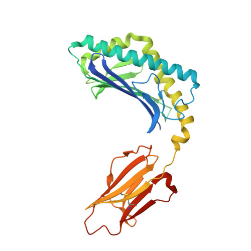
3ILQ

Structure of mCD1d with bound glycolipid BbGL-2c from Borrelia burgdorferi


Experimental Data Snapshot

  • Method: X-RAY DIFFRACTION
  • Resolution: 2.05 Å
  • R-Value Free: 0.251 
  • R-Value Work: 0.208 
  • R-Value Observed: 0.210 

Starting Model: experimental
View more details

wwPDB Validation   3D Report Full Report


Ligand Structure Quality Assessment 


This is version 2.2 of the entry. See complete history


Literature

Lipid binding orientation within CD1d affects recognition of Borrelia burgorferi antigens by NKT cells.

Wang, J.Li, Y.Kinjo, Y.Mac, T.T.Gibson, D.Painter, G.F.Kronenberg, M.Zajonc, D.M.

(2010) Proc Natl Acad Sci U S A 107: 1535-1540

  • DOI: https://doi.org/10.1073/pnas.0909479107
  • Primary Citation of Related Structures:  
    3ILP, 3ILQ

  • PubMed Abstract: 

    Invariant natural killer T cells (iNKT cells) respond to CD1d-presented glycolipids from Borrelia burgdorferi, the causative agent of Lyme disease. Although mouse and human iNKT cells respond to different antigens based on subtle differences in their fatty acids, the mechanism by which fatty acid structure determines antigenic potency is not well understood. Here we show that the mouse and human CD1d present glycolipids having different fatty acids, based in part upon a difference at a single amino acid position that is involved in positioning the sugar epitope. CD1d also can bind nonantigenic lipids, however, but unexpectedly, mouse CD1d orients the two aliphatic chains of a nonantigenic lipid rotated 180 degrees, causing a dramatic repositioning of the exposed sugar. Therefore, our data reveal the biochemical basis for the high degree of antigenic specificity of iNKT cells for certain fatty acids, and they suggest how microbes could alter fatty acid biosynthesis as an immune evasion mechanism.


  • Organizational Affiliation

    Division of Cell Biology, La Jolla Institute for Allergy and Immunology, La Jolla, CA 92037, USA.


Macromolecules
Find similar proteins by:  (by identity cutoff)  |  3D Structure
Entity ID: 1
MoleculeChains Sequence LengthOrganismDetailsImage
T-cell surface glycoprotein CD1d1A [auth C]285Mus musculusMutation(s): 0 
Gene Names: Cd1.1CD1dCd1d1
UniProt & NIH Common Fund Data Resources
Find proteins for P11609 (Mus musculus)
Explore P11609 
Go to UniProtKB:  P11609
IMPC:  MGI:107674
Entity Groups  
Sequence Clusters30% Identity50% Identity70% Identity90% Identity95% Identity100% Identity
UniProt GroupP11609
Glycosylation
Glycosylation Sites: 3Go to GlyGen: P11609-1
Sequence Annotations
Expand
  • Reference Sequence
Find similar proteins by:  (by identity cutoff)  |  3D Structure
Entity ID: 2
MoleculeChains Sequence LengthOrganismDetailsImage
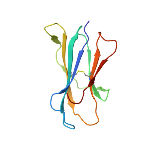
Beta-2 microglobulinB [auth D]99Mus musculusMutation(s): 0 
Gene Names: B2mbeta 2 microblobulinmCG_11606RP23-34E24.5-001
UniProt & NIH Common Fund Data Resources
Find proteins for P01887 (Mus musculus)
Explore P01887 
Go to UniProtKB:  P01887
IMPC:  MGI:88127
Entity Groups  
Sequence Clusters30% Identity50% Identity70% Identity90% Identity95% Identity100% Identity
UniProt GroupP01887
Sequence Annotations
Expand
  • Reference Sequence
Oligosaccharides

Help

Entity ID: 3
MoleculeChains Length2D Diagram Glycosylation3D Interactions
2-acetamido-2-deoxy-beta-D-glucopyranose-(1-4)-2-acetamido-2-deoxy-beta-D-glucopyranoseC [auth A]2N-Glycosylation
Glycosylation Resources
GlyTouCan:  G42666HT
GlyCosmos:  G42666HT
GlyGen:  G42666HT
Small Molecules
Ligands 2 Unique
IDChains Name / Formula / InChI Key2D Diagram3D Interactions
1O2
Query on 1O2

Download Ideal Coordinates CCD File 
F [auth C](2S)-3-(alpha-D-galactopyranosyloxy)-2-(hexadecanoyloxy)propyl (9Z)-octadec-9-enoate
C43 H80 O10
JBZBYHKCRFIXBI-BNOJPGAFSA-N
NAG
Query on NAG

Download Ideal Coordinates CCD File 
D [auth C],
E [auth C]
2-acetamido-2-deoxy-beta-D-glucopyranose
C8 H15 N O6
OVRNDRQMDRJTHS-FMDGEEDCSA-N
Binding Affinity Annotations 
IDSourceBinding Affinity
1O2 PDBBind:  3ILQ Kd: 6200 (nM) from 1 assay(s)
Experimental Data & Validation

Experimental Data

  • Method: X-RAY DIFFRACTION
  • Resolution: 2.05 Å
  • R-Value Free: 0.251 
  • R-Value Work: 0.208 
  • R-Value Observed: 0.210 
  • Space Group: P 1 21 1
Unit Cell:
Length ( Å )Angle ( ˚ )
a = 41.706α = 90
b = 97.791β = 106.94
c = 55.421γ = 90
Software Package:
Software NamePurpose
HKL-2000data collection
MOLREPphasing
REFMACrefinement
DENZOdata reduction
SCALEPACKdata scaling

Structure Validation

View Full Validation Report


Ligand Structure Quality Assessment 


Entry History 

Deposition Data

  • Released Date: 2010-01-26 
  • Deposition Author(s): Zajonc, D.M.

Revision History  (Full details and data files)

  • Version 1.0: 2010-01-26
    Type: Initial release
  • Version 1.1: 2011-07-13
    Changes: Version format compliance
  • Version 2.0: 2020-07-29
    Type: Remediation
    Reason: Carbohydrate remediation
    Changes: Advisory, Atomic model, Data collection, Database references, Derived calculations, Structure summary
  • Version 2.1: 2023-09-06
    Changes: Data collection, Database references, Refinement description, Structure summary
  • Version 2.2: 2024-10-30
    Changes: Structure summary