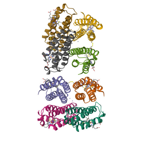
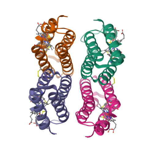
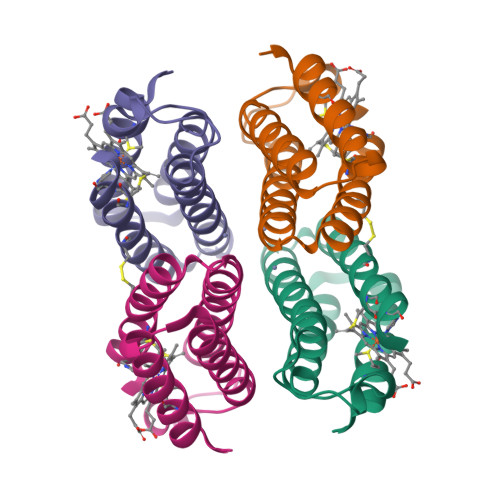
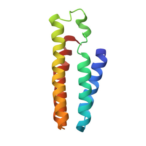
3IQ6

Crystal structure of a tetrameric Zn-bound cytochrome cb562 complex with covalently and non-covalently stabilized interfaces


Experimental Data Snapshot

  • Method: X-RAY DIFFRACTION
  • Resolution: 2.35 Å
  • R-Value Free: 0.296 
  • R-Value Work: 0.228 
  • R-Value Observed: 0.236 

Starting Model: experimental
View more details

wwPDB Validation   3D Report Full Report


Ligand Structure Quality Assessment 


This is version 1.5 of the entry. See complete history


Literature

Evolution of metal selectivity in templated protein interfaces.

Brodin, J.D.Medina-Morales, A.Ni, T.Salgado, E.N.Ambroggio, X.I.Tezcan, F.A.

(2010) J Am Chem Soc 132: 8610-8617

  • DOI: https://doi.org/10.1021/ja910844n
  • Primary Citation of Related Structures:  
    3IQ5, 3IQ6, 3M79

  • PubMed Abstract: 

    Selective binding by metalloproteins to their cognate metal ions is essential to cellular survival. How proteins originally acquired the ability to selectively bind metals and evolved a diverse array of metal-centered functions despite the availability of only a few metal-coordinating functionalities remains an open question. Using a rational design approach (Metal-Templated Interface Redesign), we describe the transformation of a monomeric electron transfer protein, cytochrome cb(562), into a tetrameric assembly ((C96)RIDC-1(4)) that stably and selectively binds Zn(2+) and displays a metal-dependent conformational change reminiscent of a signaling protein. A thorough analysis of the metal binding properties of (C96)RIDC-1(4) reveals that it can also stably harbor other divalent metals with affinities that rival (Ni(2+)) or even exceed (Cu(2+)) those of Zn(2+) on a per site basis. Nevertheless, this analysis suggests that our templating strategy simultaneously introduces an increased bias toward binding a higher number of Zn(2+) ions (four high affinity sites) versus Cu(2+) or Ni(2+) (two high affinity sites), ultimately leading to the exclusive selectivity of (C96)RIDC-1(4) for Zn(2+) over those ions. More generally, our results indicate that an initial metal-driven nucleation event followed by the formation of a stable protein architecture around the metal provides a straightforward path for generating structural and functional diversity.


  • Organizational Affiliation

    Department of Chemistry and Biochemistry, University of California, San Diego, La Jolla, California 92093-0356, USA.


Macromolecules
Find similar proteins by:  (by identity cutoff)  |  3D Structure
Entity ID: 1
MoleculeChains Sequence LengthOrganismDetailsImage
Soluble cytochrome b562
A, B, C, D, E
A, B, C, D, E, F, G, H
106Escherichia coliMutation(s): 12 
UniProt
Find proteins for P0ABE7 (Escherichia coli)
Explore P0ABE7 
Go to UniProtKB:  P0ABE7
Entity Groups  
Sequence Clusters30% Identity50% Identity70% Identity90% Identity95% Identity100% Identity
UniProt GroupP0ABE7
Sequence Annotations
Expand
  • Reference Sequence
Small Molecules
Ligands 2 Unique
IDChains Name / Formula / InChI Key2D Diagram3D Interactions
HEM
Query on HEM

Download Ideal Coordinates CCD File 
I [auth A]
K [auth B]
M [auth C]
O [auth D]
Q [auth E]
I [auth A],
K [auth B],
M [auth C],
O [auth D],
Q [auth E],
S [auth F],
U [auth G],
W [auth H]
PROTOPORPHYRIN IX CONTAINING FE
C34 H32 Fe N4 O4
KABFMIBPWCXCRK-RGGAHWMASA-L
ZN
Query on ZN

Download Ideal Coordinates CCD File 
J [auth A]
L [auth B]
N [auth C]
P [auth D]
R [auth E]
J [auth A],
L [auth B],
N [auth C],
P [auth D],
R [auth E],
T [auth F],
V [auth G],
X [auth H]
ZINC ION
Zn
PTFCDOFLOPIGGS-UHFFFAOYSA-N
Experimental Data & Validation

Experimental Data

  • Method: X-RAY DIFFRACTION
  • Resolution: 2.35 Å
  • R-Value Free: 0.296 
  • R-Value Work: 0.228 
  • R-Value Observed: 0.236 
  • Space Group: P 21 21 21
Unit Cell:
Length ( Å )Angle ( ˚ )
a = 63.265α = 90
b = 76.77β = 90
c = 177.566γ = 90
Software Package:
Software NamePurpose
DENZOdata reduction
SCALEPACKdata scaling
MOLREPphasing
CNSrefinement
PDB_EXTRACTdata extraction
Blu-Icedata collection
SCALAdata scaling

Structure Validation

View Full Validation Report


Ligand Structure Quality Assessment 


Entry History 

Deposition Data

Revision History  (Full details and data files)

  • Version 1.0: 2010-06-16
    Type: Initial release
  • Version 1.1: 2011-07-13
    Changes: Version format compliance
  • Version 1.2: 2017-11-01
    Changes: Refinement description
  • Version 1.3: 2021-10-13
    Changes: Database references, Derived calculations
  • Version 1.4: 2023-09-06
    Changes: Data collection, Refinement description
  • Version 1.5: 2024-10-16
    Changes: Structure summary