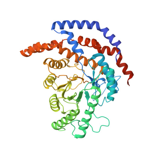
3IUH

Co2+-bound form of Pseudomonas stutzeri L-rhamnose isomerase


Experimental Data Snapshot

  • Method: X-RAY DIFFRACTION
  • Resolution: 2.00 Å
  • R-Value Free: 0.225 
  • R-Value Work: 0.183 
  • R-Value Observed: 0.183 

Starting Model: experimental
View more details

wwPDB Validation   3D Report Full Report


This is version 1.4 of the entry. See complete history


Literature

Catalytic reaction mechanism of Pseudomonas stutzeri l-rhamnose isomerase deduced from X-ray structures

Yoshida, H.Yamaji, M.Ishii, T.Izumori, K.Kamitori, S.

(2010) FEBS J 277: 1045-1057

  • DOI: https://doi.org/10.1111/j.1742-4658.2009.07548.x
  • Primary Citation of Related Structures:  
    3ITL, 3ITO, 3ITT, 3ITV, 3ITX, 3ITY, 3IUD, 3IUH, 3IUI

  • PubMed Abstract: 

    L-Rhamnose isomerase (L-RhI) catalyzes the reversible isomerization of L-rhamnose to L-rhamnulose. Pseudomonas stutzeril-RhI, with a broad substrate specificity, can catalyze not only the isomerization of L-rhamnose, but also that between D-allose and D-psicose. For the aldose-ketose isomerization by L-RhI, a metal-mediated hydride-shift mechanism has been proposed, but the catalytic mechanism is still not entirely understood. To elucidate the entire reaction mechanism, the X-ray structures of P. stutzeril-RhI in an Mn(2+)-bound form, and of two inactive mutant forms of P. stutzeril-RhI (S329K and D327N) in a complex with substrate/product, were determined. The structure of the Mn(2+)-bound enzyme indicated that the catalytic site interconverts between two forms with the displacement of the metal ion to recognize both pyranose and furanose ring substrates. Solving the structures of S329K-substrates allowed us to examine the metal-mediated hydride-shift mechanism of L-RhI in detail. The structural analysis of D327N-substrates and additional modeling revealed Asp327 to be responsible for the ring opening of furanose, and a water molecule coordinating with the metal ion to be involved in the ring opening of pyranose.


  • Organizational Affiliation

    Life Science Research Center and Faculty of Medicine, Kagawa University, Japan.


Macromolecules
Find similar proteins by:  (by identity cutoff)  |  3D Structure
Entity ID: 1
MoleculeChains Sequence LengthOrganismDetailsImage
L-rhamnose isomerase
A, B, C, D
438Stutzerimonas stutzeriMutation(s): 1 
EC: 5.3.1.14
UniProt
Find proteins for Q75WH8 (Stutzerimonas stutzeri)
Explore Q75WH8 
Go to UniProtKB:  Q75WH8
Entity Groups  
Sequence Clusters30% Identity50% Identity70% Identity90% Identity95% Identity100% Identity
UniProt GroupQ75WH8
Sequence Annotations
Expand
  • Reference Sequence
Experimental Data & Validation

Experimental Data

  • Method: X-RAY DIFFRACTION
  • Resolution: 2.00 Å
  • R-Value Free: 0.225 
  • R-Value Work: 0.183 
  • R-Value Observed: 0.183 
  • Space Group: P 1 21 1
Unit Cell:
Length ( Å )Angle ( ˚ )
a = 78.072α = 90
b = 104.96β = 108.23
c = 113.369γ = 90
Software Package:
Software NamePurpose
ADSCdata collection
MOLREPphasing
CNSrefinement
HKL-2000data reduction
HKL-2000data scaling

Structure Validation

View Full Validation Report


Entry History 

Deposition Data

Revision History  (Full details and data files)

  • Version 1.0: 2010-02-02
    Type: Initial release
  • Version 1.1: 2011-07-13
    Changes: Version format compliance
  • Version 1.2: 2019-11-20
    Changes: Database references, Derived calculations
  • Version 1.3: 2021-11-10
    Changes: Database references, Derived calculations
  • Version 1.4: 2023-11-01
    Changes: Data collection, Refinement description