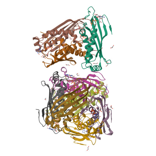
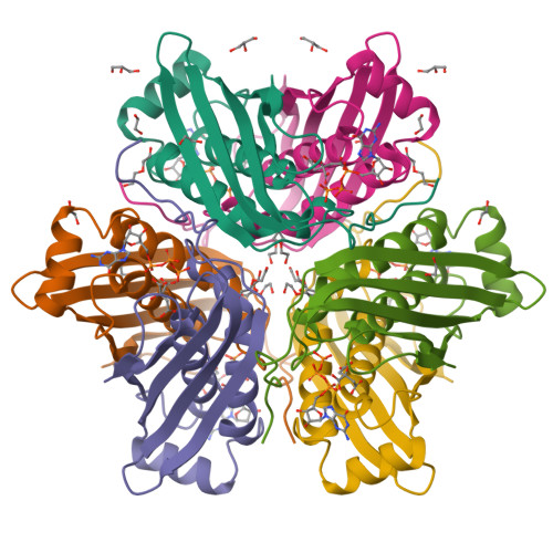
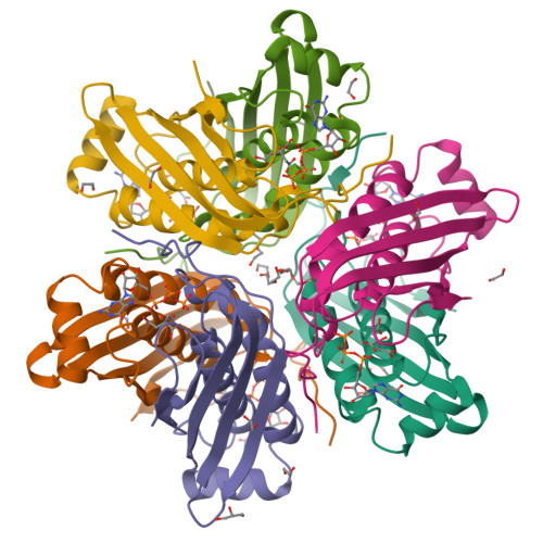
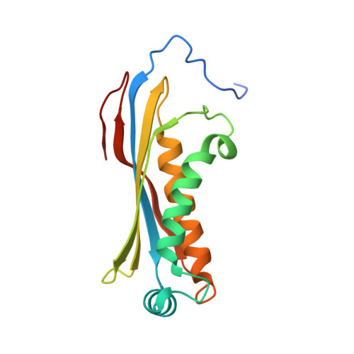
3JQM | pdb_00003jqm

Binding of 5'-GTP to molybdenum cofactor biosynthesis protein MoaC from Thermus theromophilus HB8


Experimental Data Snapshot

  • Method: X-RAY DIFFRACTION
  • Resolution: 2.50 Å
  • R-Value Free: 
    0.270 (Depositor), 0.260 (DCC) 
  • R-Value Work: 
    0.202 (Depositor), 0.200 (DCC) 
  • R-Value Observed: 
    0.202 (Depositor) 

Starting Model: experimental
View more details

wwPDB Validation   3D Report Full Report


Ligand Structure Quality Assessment 

Created with Raphaël 2.3.0Worse 01 BetterLigand structure goodness of fit to experimental dataBest fitted GTPClick on this verticalbar to view details

This is version 1.3 of the entry. See complete history


Literature

Structures of apo and GTP-bound molybdenum cofactor biosynthesis protein MoaC from Thermus thermophilus HB8

Kanaujia, S.P.Jeyakanthan, J.Nakagawa, N.Balasubramaniam, S.Shinkai, A.Kuramitsu, S.Yokoyama, S.Sekar, K.

(2010) Acta Crystallogr D Biol Crystallogr 66: 821-833

  • DOI: https://doi.org/10.1107/S0907444910019074
  • Primary Citation of Related Structures:  
    3JQJ, 3JQK, 3JQM

  • PubMed Abstract: 

    The first step in the molybdenum cofactor (Moco) biosynthesis pathway involves the conversion of guanosine triphosphate (GTP) to precursor Z by two proteins (MoaA and MoaC). MoaA belongs to the S-adenosylmethionine-dependent radical enzyme superfamily and is believed to generate protein and/or substrate radicals by reductive cleavage of S-adenosylmethionine using an Fe-S cluster. MoaC has been suggested to catalyze the release of pyrophosphate and the formation of the cyclic phosphate of precursor Z. However, structural evidence showing the binding of a substrate-like molecule to MoaC is not available. Here, apo and GTP-bound crystal structures of MoaC from Thermus thermophilus HB8 are reported. Furthermore, isothermal titration calorimetry experiments have been carried out in order to obtain thermodynamic parameters for the protein-ligand interactions. In addition, molecular-dynamics (MD) simulations have been carried out on the protein-ligand complex of known structure and on models of relevant complexes for which X-ray structures are not available. The biophysical, structural and MD results reveal the residues that are involved in substrate binding and help in speculating upon a possible mechanism.


  • Organizational Affiliation

    Bioinformatics Centre (Centre of Excellence in Structural Biology and Bio-computing), Indian Institute of Science, Bangalore, India.


Macromolecules
Find similar proteins by:  (by identity cutoff)  |  3D Structure
Entity ID: 1
MoleculeChains Sequence LengthOrganismDetailsImage
Molybdenum cofactor biosynthesis protein C
A, B, C, D, E
157Thermus thermophilus HB8Mutation(s): 0 
Gene Names: TTHA1789
EC: 4.6.1.17
UniProt
Find proteins for Q5SHE1 (Thermus thermophilus (strain ATCC 27634 / DSM 579 / HB8))
Explore Q5SHE1 
Go to UniProtKB:  Q5SHE1
Entity Groups  
Sequence Clusters30% Identity50% Identity70% Identity90% Identity95% Identity100% Identity
UniProt GroupQ5SHE1
Sequence Annotations
Expand
  • Reference Sequence
Small Molecules
Ligands 5 Unique
IDChains Name / Formula / InChI Key2D Diagram3D Interactions
GTP
Query on GTP

Download Ideal Coordinates CCD File 
BB [auth I]
CA [auth D]
GA [auth E]
J [auth A]
JA [auth F]
GUANOSINE-5'-TRIPHOSPHATE
C10 H16 N5 O14 P3
XKMLYUALXHKNFT-UUOKFMHZSA-N
FLC
Query on FLC

Download Ideal Coordinates CCD File 
CB [auth I]
DA [auth D]
HA [auth E]
K [auth A]
KA [auth F]
CITRATE ANION
C6 H5 O7
KRKNYBCHXYNGOX-UHFFFAOYSA-K
PEG
Query on PEG

Download Ideal Coordinates CCD File 
AB [auth H]
BA [auth C]
IA [auth E]
OA [auth F]
P [auth A]
DI(HYDROXYETHYL)ETHER
C4 H10 O3
MTHSVFCYNBDYFN-UHFFFAOYSA-N
GOL
Query on GOL

Download Ideal Coordinates CCD File 
L [auth A],
LA [auth F],
M [auth A],
XA [auth H],
Z [auth C]
GLYCEROL
C3 H8 O3
PEDCQBHIVMGVHV-UHFFFAOYSA-N
EDO
Query on EDO

Download Ideal Coordinates CCD File 
AA [auth C]
DB [auth I]
EA [auth D]
EB [auth I]
FA [auth D]
1,2-ETHANEDIOL
C2 H6 O2
LYCAIKOWRPUZTN-UHFFFAOYSA-N
Experimental Data & Validation

Experimental Data

  • Method: X-RAY DIFFRACTION
  • Resolution: 2.50 Å
  • R-Value Free:  0.270 (Depositor), 0.260 (DCC) 
  • R-Value Work:  0.202 (Depositor), 0.200 (DCC) 
  • R-Value Observed: 0.202 (Depositor) 
Space Group: C 2 2 21
Unit Cell:
Length ( Å )Angle ( ˚ )
a = 69.929α = 90
b = 111.567β = 90
c = 311.423γ = 90
Software Package:
Software NamePurpose
HKL-2000data collection
PHASERphasing
CNSrefinement
HKL-2000data reduction
HKL-2000data scaling

Structure Validation

View Full Validation Report


Ligand Structure Quality Assessment 

Created with Raphaël 2.3.0Worse 01 BetterLigand structure goodness of fit to experimental dataBest fitted GTPClick on this verticalbar to view details

Entry History 

Revision History  (Full details and data files)

  • Version 1.0: 2010-06-30
    Type: Initial release
  • Version 1.1: 2011-07-13
    Changes: Version format compliance
  • Version 1.2: 2012-04-11
    Changes: Database references
  • Version 1.3: 2023-11-01
    Changes: Data collection, Database references, Derived calculations, Refinement description