3LBW

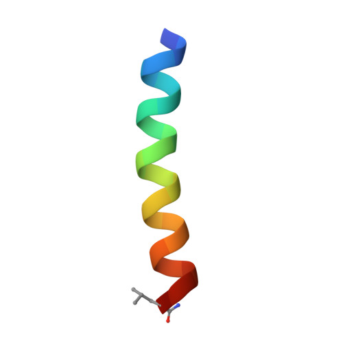
High resolution crystal structure of transmembrane domain of M2


Experimental Data Snapshot

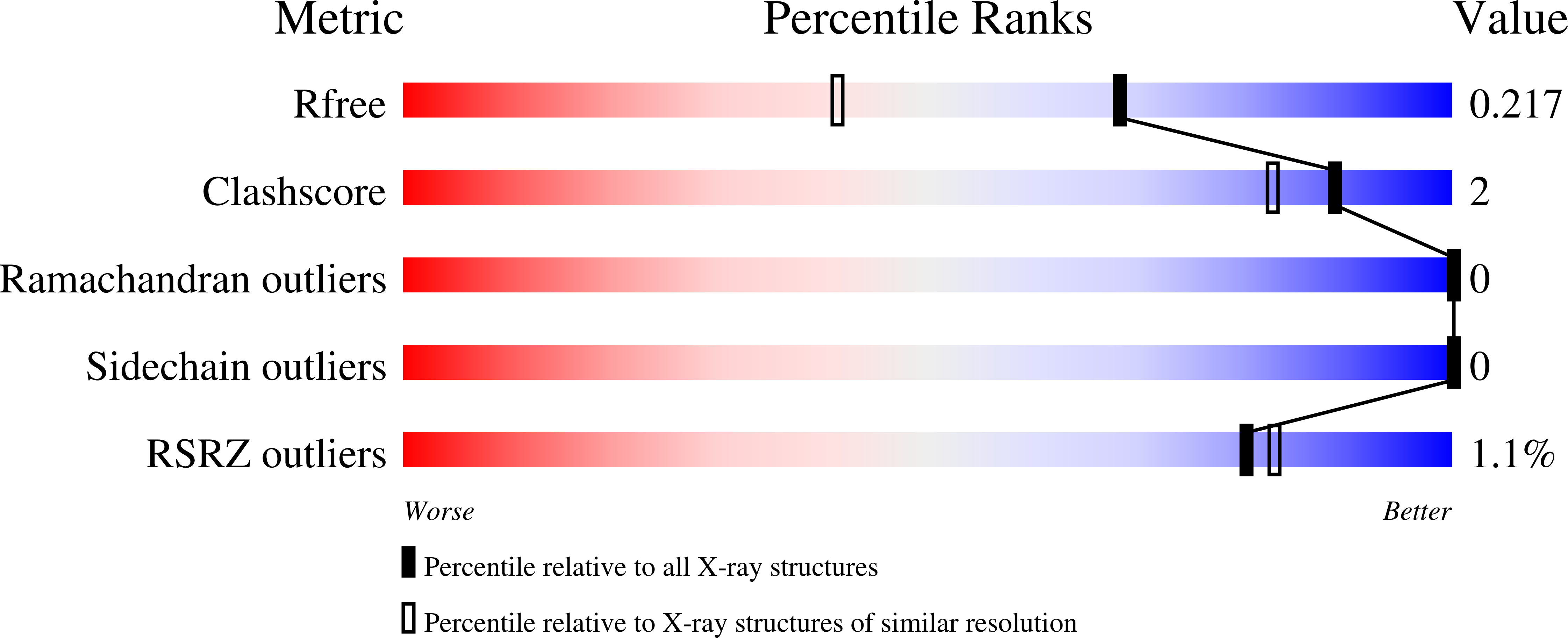
  • Method: X-RAY DIFFRACTION
  • Resolution: 1.65 Å
  • R-Value Free: 0.205 
  • R-Value Work: 0.196 
  • R-Value Observed: 0.197 

Starting Model: experimental
View more details

wwPDB Validation   3D Report Full Report


Ligand Structure Quality Assessment 


This is version 1.5 of the entry. See complete history


Literature

Structure and mechanism of proton transport through the transmembrane tetrameric M2 protein bundle of the influenza A virus.

Acharya, R.Carnevale, V.Fiorin, G.Levine, B.G.Polishchuk, A.L.Balannik, V.Samish, I.Lamb, R.A.Pinto, L.H.DeGrado, W.F.Klein, M.L.

(2010) Proc Natl Acad Sci U S A 107: 15075-15080

  • DOI: https://doi.org/10.1073/pnas.1007071107
  • Primary Citation of Related Structures:  
    3LBW

  • PubMed Abstract: 

    The M2 proton channel from influenza A virus is an essential protein that mediates transport of protons across the viral envelope. This protein has a single transmembrane helix, which tetramerizes into the active channel. At the heart of the conduction mechanism is the exchange of protons between the His37 imidazole moieties of M2 and waters confined to the M2 bundle interior. Protons are conducted as the total charge of the four His37 side chains passes through 2(+) and 3(+) with a pK(a) near 6. A 1.65 A resolution X-ray structure of the transmembrane protein (residues 25-46), crystallized at pH 6.5, reveals a pore that is lined by alternating layers of sidechains and well-ordered water clusters, which offer a pathway for proton conduction. The His37 residues form a box-like structure, bounded on either side by water clusters with well-ordered oxygen atoms at close distance. The conformation of the protein, which is intermediate between structures previously solved at higher and lower pH, suggests a mechanism by which conformational changes might facilitate asymmetric diffusion through the channel in the presence of a proton gradient. Moreover, protons diffusing through the channel need not be localized to a single His37 imidazole, but instead may be delocalized over the entire His-box and associated water clusters. Thus, the new crystal structure provides a possible unification of the discrete site versus continuum conduction models.


  • Organizational Affiliation

    Department of Biochemistry and Biophysics, School of Medicine, University of Pennsylvania, Philadelphia, PA 19104-6059.


Macromolecules

Find similar proteins by:  Sequence   |   3D Structure  

Entity ID: 1
MoleculeChains Sequence LengthOrganismDetailsImage
M2 protein
A, B, C, D
23H3N2 subtypeMutation(s): 2 
Membrane Entity: Yes 
UniProt
Find proteins for O70632 (Influenza A virus (strain A/Hong Kong/156/1997 H5N1 genotype Gs/Gd))
Explore O70632 
Go to UniProtKB:  O70632
Entity Groups  
Sequence Clusters30% Identity50% Identity70% Identity90% Identity95% Identity100% Identity
UniProt GroupO70632
Sequence Annotations
Expand
  • Reference Sequence
Small Molecules
Ligands 3 Unique
IDChains Name / Formula / InChI Key2D Diagram3D Interactions
Z82
Query on Z82

Download Ideal Coordinates CCD File 
E [auth A],
G [auth B],
I [auth C],
L [auth D]
4-bromobenzoic acid
C7 H5 Br O2
TUXYZHVUPGXXQG-UHFFFAOYSA-N
XYL
Query on XYL

Download Ideal Coordinates CCD File 
H [auth B]Xylitol
C5 H12 O5
HEBKCHPVOIAQTA-SCDXWVJYSA-N
PEG
Query on PEG

Download Ideal Coordinates CCD File 
F [auth A],
J [auth C],
K [auth C]
DI(HYDROXYETHYL)ETHER
C4 H10 O3
MTHSVFCYNBDYFN-UHFFFAOYSA-N
Experimental Data & Validation

Experimental Data

  • Method: X-RAY DIFFRACTION
  • Resolution: 1.65 Å
  • R-Value Free: 0.205 
  • R-Value Work: 0.196 
  • R-Value Observed: 0.197 
  • Space Group: C 2 2 21
Unit Cell:
Length ( Å )Angle ( ˚ )
a = 48.665α = 90
b = 79.091β = 90
c = 48.559γ = 90
Software Package:
Software NamePurpose
CBASSdata collection
PHASERphasing
REFMACrefinement
MOSFLMdata reduction
SCALAdata scaling

Structure Validation

View Full Validation Report


Ligand Structure Quality Assessment 


Entry History 

Deposition Data

Revision History  (Full details and data files)

  • Version 1.0: 2010-07-28
    Type: Initial release
  • Version 1.1: 2011-07-13
    Changes: Version format compliance
  • Version 1.2: 2011-08-10
    Changes: Database references
  • Version 1.3: 2020-07-29
    Type: Remediation
    Reason: Carbohydrate remediation
    Changes: Database references, Derived calculations, Structure summary
  • Version 1.4: 2021-10-13
    Changes: Database references, Structure summary
  • Version 1.5: 2023-09-06
    Changes: Data collection, Refinement description