3MYW

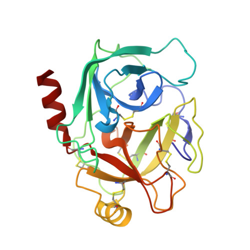
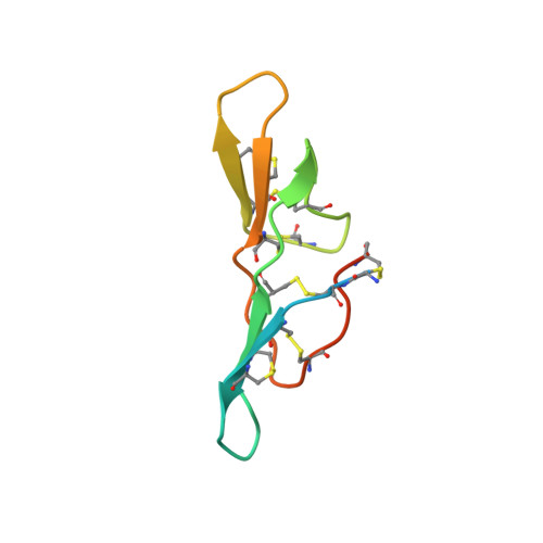
The Bowman-Birk type inhibitor from mung bean in ternary complex with porcine trypsin


Experimental Data Snapshot

  • Method: X-RAY DIFFRACTION
  • Resolution: 2.50 Å
  • R-Value Observed: 0.179 

Starting Model: experimental
View more details

wwPDB Validation   3D Report Full Report


This is version 1.5 of the entry. See complete history


Literature

The 0.25-nm X-ray structure of the Bowman-Birk-type inhibitor from mung bean in ternary complex with porcine trypsin.

Lin, G.Bode, W.Huber, R.Chi, C.Engh, R.A.

(1993) Eur J Biochem 212: 549-555

  • DOI: https://doi.org/10.1111/j.1432-1033.1993.tb17692.x
  • Primary Citation of Related Structures:  
    3MYW

  • PubMed Abstract: 

    The structure of the Bowman-Birk-type inhibitor from mung bean Phaseolus aureus has been determined in ternary complex with porcine trypsin. The complex formed crystals of the trigonal space group P3(1)21 which diffracted to a resolution of 250 pm. Each of the two mung bean protease reactive sites is bound to trypsin according to the standard mechanism for serine proteinase inhibition. The binding loops thereby adopt the canonical conformation for the standard mechanism; however, the sub-van der Waals contact between the active-site serine O gamma (195) and the P1 carbonyl carbon of both loops is significantly smaller (210 pm) than hitherto observed, with continuous electron density connecting the two atoms. The inhibitor is formed by two double-stranded antiparallel beta-sheets, which are connected into a moderately twisted beta-sheet by a network of hydrogen bonds involving main-chain atoms and two water molecules. All contacts with neighbors in the crystal lattice occur between trypsin molecules. This apparently gives rise to an unusual form of disorder where the complexes pack in two orientations Ta:MaMb:Tb and Tb:MbMa:Ta (Ta, Tb = trypsin, Ma = mung bean loop I, Mb = mung bean loop II), such that the asymmetric unit consists of the ternary complex in two orientations, each with half occupancy. This is nearly equivalent to an asymmetric unit which has one trypsin molecule with full occupancy and one mung bean inhibitor with half occupancy and a crystallographic twofold symmetry axis through its center. Because of the approximate twofold symmetry of the inhibitor itself, however, the electron density was interpretable for most of the inhibitor (17 residues at the termini were not resolved) and shows evidence of its double orientation.


  • Organizational Affiliation

    Shanghai Institute of Biochemistry, People's Republic of China.


Macromolecules
Find similar proteins by:  (by identity cutoff)  |  3D Structure
Entity ID: 1
MoleculeChains Sequence LengthOrganismDetailsImage
TrypsinA,
C [auth B]
223Sus scrofaMutation(s): 1 
EC: 3.4.21.4
UniProt
Find proteins for P00761 (Sus scrofa)
Explore P00761 
Go to UniProtKB:  P00761
Entity Groups  
Sequence Clusters30% Identity50% Identity70% Identity90% Identity95% Identity100% Identity
UniProt GroupP00761
Sequence Annotations
Expand
  • Reference Sequence
Find similar proteins by:  (by identity cutoff)  |  3D Structure
Entity ID: 2
MoleculeChains Sequence LengthOrganismDetailsImage
Bowman-Birk type trypsin inhibitorB [auth I]72Vigna radiata var. radiataMutation(s): 0 
UniProt
Find proteins for P01062 (Vigna radiata var. radiata)
Explore P01062 
Go to UniProtKB:  P01062
Entity Groups  
Sequence Clusters30% Identity50% Identity70% Identity90% Identity95% Identity100% Identity
UniProt GroupP01062
Sequence Annotations
Expand
  • Reference Sequence
Experimental Data & Validation

Experimental Data

  • Method: X-RAY DIFFRACTION
  • Resolution: 2.50 Å
  • R-Value Observed: 0.179 
  • Space Group: P 31
Unit Cell:
Length ( Å )Angle ( ˚ )
a = 62.46α = 90
b = 62.46β = 90
c = 160.01γ = 120
Software Package:
Software NamePurpose
MADNESSdata collection
PROTEINmodel building
X-PLORrefinement
PROTEINdata reduction
PROTEINdata scaling
PROTEINphasing

Structure Validation

View Full Validation Report


Entry History 

Deposition Data

Revision History  (Full details and data files)

  • Version 1.0: 2010-12-29
    Type: Initial release
  • Version 1.1: 2011-07-13
    Changes: Version format compliance
  • Version 1.2: 2017-11-08
    Changes: Advisory, Refinement description
  • Version 1.3: 2018-04-18
    Changes: Data collection
  • Version 1.4: 2023-09-06
    Changes: Advisory, Data collection, Database references, Derived calculations, Refinement description
  • Version 1.5: 2024-11-06
    Changes: Structure summary