3OGS

Complex structure of beta-galactosidase from Trichoderma reesei with IPTG


Experimental Data Snapshot

  • Method: X-RAY DIFFRACTION
  • Resolution: 1.75 Å
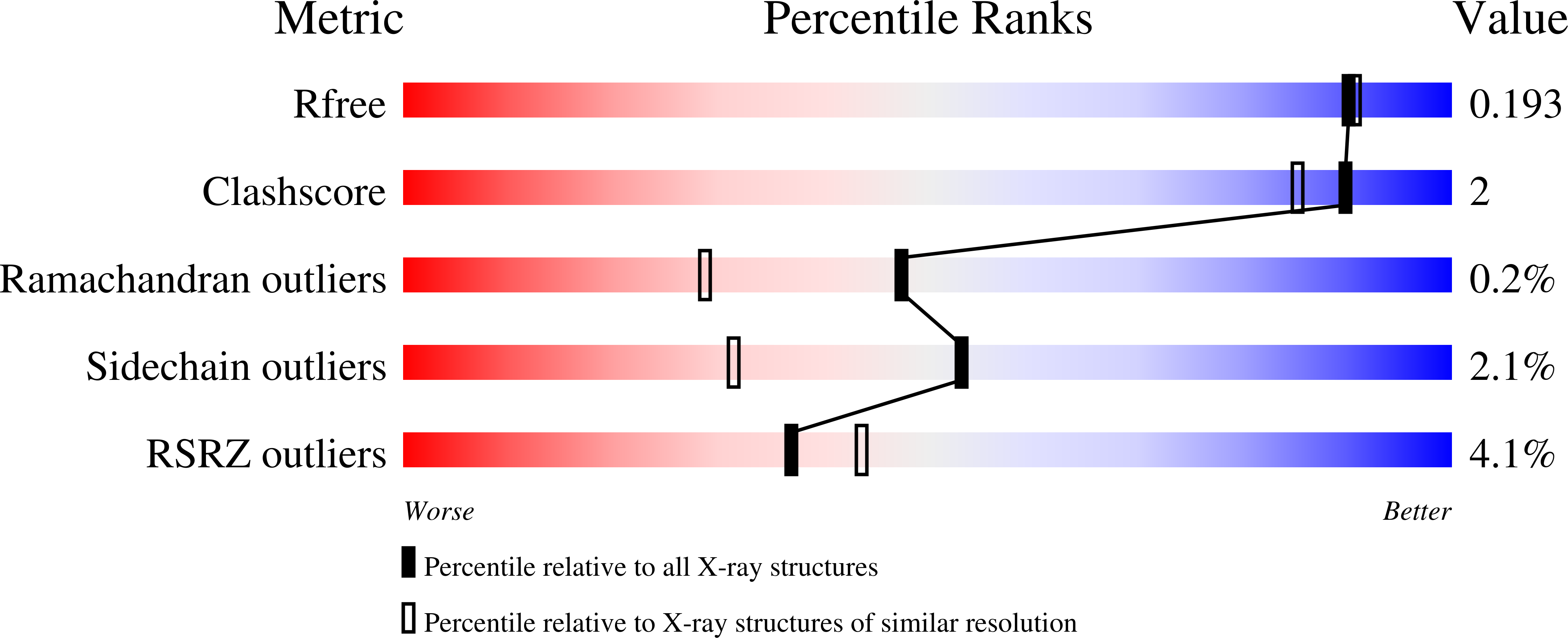
  • R-Value Free: 0.200 
  • R-Value Work: 0.145 
  • R-Value Observed: 0.148 

Starting Model: experimental
View more details

wwPDB Validation   3D Report Full Report


Ligand Structure Quality Assessment 


This is version 2.1 of the entry. See complete history


Literature

Crystal structures of Trichoderma reesei beta-galactosidase reveal conformational changes in the active site

Maksimainen, M.Hakulinen, N.Kallio, J.M.Timoharju, T.Turunen, O.Rouvinen, J.

(2011) J Struct Biol 174: 156-163

  • DOI: https://doi.org/10.1016/j.jsb.2010.11.024
  • Primary Citation of Related Structures:  
    3OG2, 3OGR, 3OGS, 3OGV

  • PubMed Abstract: 

    We have determined the crystal structure of Trichoderma reesei (Hypocrea jecorina) β-galactosidase (Tr-β-gal) at a 1.2Å resolution and its complex structures with galactose, IPTG and PETG at 1.5, 1.75 and 1.4Å resolutions, respectively. Tr-β-gal is a potential enzyme for lactose hydrolysis in the dairy industry and belongs to family 35 of the glycoside hydrolases (GH-35). The high resolution crystal structures of this six-domain enzyme revealed interesting features about the structure of Tr-β-gal. We discovered conformational changes in the two loop regions in the active site, implicating a conformational selection-mechanism for the enzyme. In addition, the Glu200, an acid/base catalyst showed two different conformations which undoubtedly affect the pK(a) value of this residue and the catalytic mechanism. The electron density showed extensive glycosylation, suggesting a structure stabilizing role for glycans. The longest glycan showed an electron density that extends to the eighth monosaccharide unit in the extended chain. The Tr-β-gal structure also showed a well-ordered structure for a unique octaserine motif on the surface loop of the fifth domain.


  • Organizational Affiliation

    Department of Chemistry, University of Eastern Finland, P.O. Box 111, FIN-80101 Joensuu, Finland.


Macromolecules
Find similar proteins by:  (by identity cutoff)  |  3D Structure
Entity ID: 1
MoleculeChains Sequence LengthOrganismDetailsImage
Beta-galactosidase1,003Trichoderma reeseiMutation(s): 0 
EC: 3.2.1.23
UniProt
Find proteins for Q70SY0 (Hypocrea jecorina)
Explore Q70SY0 
Go to UniProtKB:  Q70SY0
Entity Groups  
Sequence Clusters30% Identity50% Identity70% Identity90% Identity95% Identity100% Identity
UniProt GroupQ70SY0
Glycosylation
Glycosylation Sites: 5
Sequence Annotations
Expand
  • Reference Sequence
Oligosaccharides

Help

Entity ID: 2
MoleculeChains Length2D Diagram Glycosylation3D Interactions
alpha-D-mannopyranose-(1-2)-alpha-D-mannopyranose-(1-3)-alpha-D-mannopyranose-(1-6)-[alpha-D-mannopyranose-(1-3)]beta-D-mannopyranose-(1-4)-2-acetamido-2-deoxy-beta-D-glucopyranose-(1-4)-2-acetamido-2-deoxy-beta-D-glucopyranose
B
7N-Glycosylation
Glycosylation Resources
GlyTouCan:  G16404YH
GlyCosmos:  G16404YH
GlyGen:  G16404YH
Entity ID: 3
MoleculeChains Length2D Diagram Glycosylation3D Interactions
alpha-D-glucopyranose-(1-3)-alpha-D-mannopyranose-(1-2)-alpha-D-mannopyranose-(1-2)-alpha-D-mannopyranose-(1-3)-[alpha-D-mannopyranose-(1-3)-alpha-D-mannopyranose-(1-6)]beta-D-mannopyranose-(1-4)-2-acetamido-2-deoxy-beta-D-glucopyranose-(1-4)-2-acetamido-2-deoxy-beta-D-glucopyranose
C
9N-Glycosylation
Glycosylation Resources
GlyTouCan:  G27171ZS
GlyCosmos:  G27171ZS
GlyGen:  G27171ZS
Entity ID: 4
MoleculeChains Length2D Diagram Glycosylation3D Interactions
2-acetamido-2-deoxy-beta-D-glucopyranose-(1-4)-2-acetamido-2-deoxy-beta-D-glucopyranose
D
2N-Glycosylation
Glycosylation Resources
GlyTouCan:  G42666HT
GlyCosmos:  G42666HT
GlyGen:  G42666HT
Experimental Data & Validation

Experimental Data

  • Method: X-RAY DIFFRACTION
  • Resolution: 1.75 Å
  • R-Value Free: 0.200 
  • R-Value Work: 0.145 
  • R-Value Observed: 0.148 
  • Space Group: P 1
Unit Cell:
Length ( Å )Angle ( ˚ )
a = 69.5α = 108.5
b = 70.3β = 97.8
c = 82.4γ = 114.4
Software Package:
Software NamePurpose
DNAdata collection
MOLREPphasing
REFMACrefinement
XDSdata reduction
XSCALEdata scaling

Structure Validation

View Full Validation Report


Ligand Structure Quality Assessment 


Entry History 

Deposition Data

Revision History  (Full details and data files)

  • Version 1.0: 2011-03-16
    Type: Initial release
  • Version 1.1: 2011-07-13
    Changes: Version format compliance
  • Version 2.0: 2020-07-29
    Type: Remediation
    Reason: Carbohydrate remediation
    Changes: Atomic model, Data collection, Derived calculations, Structure summary
  • Version 2.1: 2023-11-01
    Changes: Data collection, Database references, Refinement description, Structure summary