3S1W

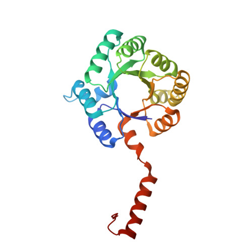
Transaldolase variant Lys86Ala from Thermoplasma acidophilum in complex with glycerol and citrate


Experimental Data Snapshot

  • Method: X-RAY DIFFRACTION
  • Resolution: 1.80 Å
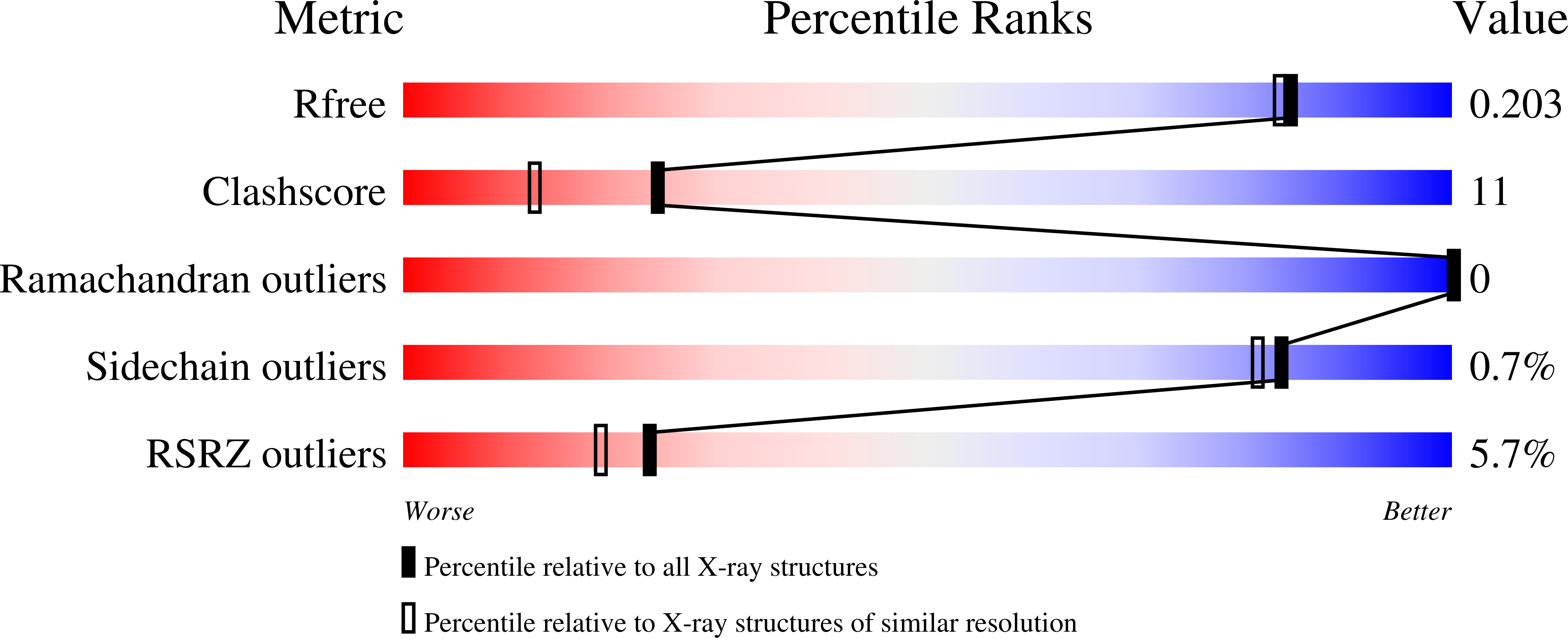
  • R-Value Free: 0.208 
  • R-Value Work: 0.176 
  • R-Value Observed: 0.178 

Starting Model: experimental
View more details

wwPDB Validation   3D Report Full Report


This is version 1.4 of the entry. See complete history


Literature

Twisted Schiff base intermediates and substrate locale revise transaldolase mechanism.

Lehwess-Litzmann, A.Neumann, P.Parthier, C.Ludtke, S.Golbik, R.Ficner, R.Tittmann, K.

(2011) Nat Chem Biol 7: 678-684

  • DOI: https://doi.org/10.1038/nchembio.633
  • Primary Citation of Related Structures:  
    3S0C, 3S1U, 3S1V, 3S1W, 3S1X

  • PubMed Abstract: 

    We examined the catalytic cycle of transaldolase (TAL) from Thermoplasma acidophilum by cryocrystallography and were able to structurally characterize--for the first time, to our knowledge--different genuine TAL reaction intermediates. These include the Schiff base adducts formed between the catalytic lysine and the donor ketose substrates fructose-6-phosphate and sedoheptulose-7-phosphate as well as the Michaelis complex with acceptor aldose erythrose-4-phosphate. These structural snapshots necessitate a revision of the accepted reaction mechanism with respect to functional roles of active site residues, and they further reveal fundamental insights into the general structural features of enzymatic Schiff base intermediates and the role of conformational dynamics in enzyme catalysis, substrate binding and discrimination. A nonplanar arrangement of the substituents around the Schiff base double bond was observed, suggesting that a structurally encoded reactant-state destabilization is a driving force of catalysis. Protein dynamics and the intrinsic hydrogen-bonding pattern appear to be crucial for selective recognition and binding of ketose as first substrate.


  • Organizational Affiliation

    Albrecht-von-Haller-Institut and Göttingen Center for Molecular Biosciences, Georg-August-Universität Göttingen, Göttingen, Germany.


Macromolecules
Find similar proteins by:  (by identity cutoff)  |  3D Structure
Entity ID: 1
MoleculeChains Sequence LengthOrganismDetailsImage
Probable transaldolase
A, B, C, D, E
223Thermoplasma acidophilum DSM 1728Mutation(s): 1 
Gene Names: Ta0616tal
EC: 2.2.1.2
UniProt
Find proteins for Q9HKI3 (Thermoplasma acidophilum (strain ATCC 25905 / DSM 1728 / JCM 9062 / NBRC 15155 / AMRC-C165))
Go to UniProtKB:  Q9HKI3
Sequence Annotations
Expand
  • Reference Sequence
Small Molecules
Ligands 2 Unique
IDChains Name / Formula / InChI Key2D Diagram3D Interactions
FLC
Query on FLC

Download Ideal Coordinates CCD File 
G [auth A],
I [auth B],
K [auth C],
M [auth D],
O [auth E]
CITRATE ANION
C6 H5 O7
KRKNYBCHXYNGOX-UHFFFAOYSA-K
GOL
Query on GOL

Download Ideal Coordinates CCD File 
F [auth A],
H [auth B],
J [auth C],
L [auth D],
N [auth E]
GLYCEROL
C3 H8 O3
PEDCQBHIVMGVHV-UHFFFAOYSA-N
Experimental Data & Validation

Experimental Data

  • Method: X-RAY DIFFRACTION
  • Resolution: 1.80 Å
  • R-Value Free: 0.208 
  • R-Value Work: 0.176 
  • R-Value Observed: 0.178 
  • Space Group: C 2 2 21
Unit Cell:
Length ( Å )Angle ( ˚ )
a = 145.08α = 90
b = 168.67β = 90
c = 97.92γ = 90
Software Package:
Software NamePurpose
XSCALEdata scaling
PHENIXrefinement
PDB_EXTRACTdata extraction
MAR345dtbdata collection
XDSdata reduction
PHENIXphasing

Structure Validation

View Full Validation Report


Entry History 

Deposition Data

Revision History  (Full details and data files)

  • Version 1.0: 2011-08-24
    Type: Initial release
  • Version 1.1: 2011-09-07
    Changes: Database references
  • Version 1.2: 2011-10-05
    Changes: Database references
  • Version 1.3: 2019-09-04
    Changes: Data collection
  • Version 1.4: 2023-09-13
    Changes: Data collection, Database references, Derived calculations, Refinement description