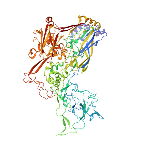
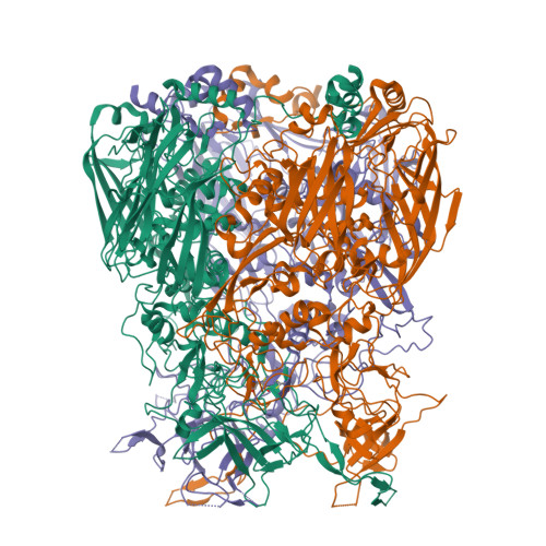
crystal structure of Adenovirus serotype 5 hexon at 1.6A resolution
Zhu, Y., Roszak, A.W., Isaacs, N.W., McVey, J.H., Nicklin, S.A., Baker, A.H.To be published.
Experimental Data Snapshot
Starting Model: experimental
View more details
wwPDB Validation 3D Report Full Report

Entity ID: 1 | |||||
|---|---|---|---|---|---|
| Molecule | Chains | Sequence Length | Organism | Details | Image |
| Hexon protein | 951 | Human adenovirus 5 | Mutation(s): 0 | 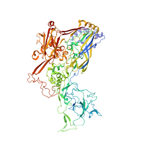 | |
UniProt | |||||
Find proteins for P04133 (Human adenovirus C serotype 5) Explore P04133 Go to UniProtKB: P04133 | |||||
Entity Groups | |||||
| Sequence Clusters | 30% Identity50% Identity70% Identity90% Identity95% Identity100% Identity | ||||
| UniProt Group | P04133 | ||||
Sequence AnnotationsExpand | |||||
| |||||
| Length ( Å ) | Angle ( ˚ ) |
|---|---|
| a = 149.15 | α = 90 |
| b = 149.15 | β = 90 |
| c = 126.69 | γ = 120 |
| Software Name | Purpose |
|---|---|
| PHASER | phasing |
| REFMAC | refinement |
| d*TREK | data reduction |
| d*TREK | data scaling |
RCSB PDB is hosted by

RCSB PDB is a member of the