S-SAD phasing study of death receptor 6 and its solution conformation revealed by SAXS
Ru, H., Zhao, L.X., Ding, W., Jiao, L.Y., Shaw, N., Liang, W., Zhang, L.G., Hung, L.W., Matsugaki, N., Wakatsuki, S., Liu, Z.J.(2012) Acta Crystallogr D Biol Crystallogr 68: 521-530
- PubMed: 22525750
- DOI: https://doi.org/10.1107/S0907444912004490
- Primary Citation of Related Structures:
3U3P, 3U3Q, 3U3S, 3U3T, 3U3V - PubMed Abstract:
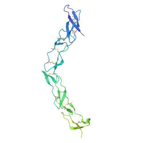
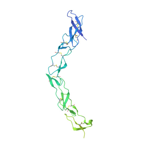
A subset of tumour necrosis factor receptor (TNFR) superfamily members contain death domains in their cytoplasmic tails. Death receptor 6 (DR6) is one such member and can trigger apoptosis upon the binding of a ligand by its cysteine-rich domains (CRDs). The crystal structure of the ectodomain (amino acids 1-348) of human death receptor 6 (DR6) encompassing the CRD region was phased using the anomalous signal from S atoms. In order to explore the feasibility of S-SAD phasing at longer wavelengths (beyond 2.5 Å), a comparative study was performed on data collected at wavelengths of 2.0 and 2.7 Å. In spite of sub-optimal experimental conditions, the 2.7 Å wavelength used for data collection showed potential for S-SAD phasing. The results showed that the R(ano)/R(p.i.m.) ratio is a good indicator for monitoring the anomalous data quality when the anomalous signal is relatively strong, while d''/sig(d'') calculated by SHELXC is a more sensitive and stable indicator applicable for grading a wider range of anomalous data qualities. The use of the `parameter-space screening method' for S-SAD phasing resulted in solutions for data sets that failed during manual attempts. SAXS measurements on the ectodomain suggested that a dimer defines the minimal physical unit of an unliganded DR6 molecule in solution.
Organizational Affiliation:
National Laboratory of Biomacromolecules, Institute of Biophysics, Chinese Academy of Sciences, Beijing 100101, People's Republic of China.