3V5U | pdb_00003v5u

Structure of Sodium/Calcium Exchanger from Methanocaldococcus jannaschii DSM 2661


Experimental Data Snapshot

  • Method: X-RAY DIFFRACTION
  • Resolution: 1.90 Å
  • R-Value Free: 
    0.210 (Depositor), 0.210 (DCC) 
  • R-Value Work: 
    0.185 (Depositor), 0.180 (DCC) 
  • R-Value Observed: 
    0.187 (Depositor) 

wwPDB Validation   3D Report Full Report


Ligand Structure Quality Assessment 

Created with Raphaël 2.3.0Worse 01 BetterLigand structure goodness of fit to experimental dataBest fitted OLCClick on this verticalbar to view detailsBest fitted MYSClick on this verticalbar to view details

This is version 1.1 of the entry. See complete history


Literature

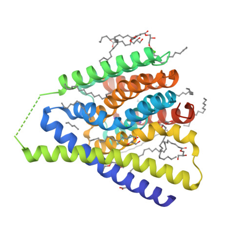
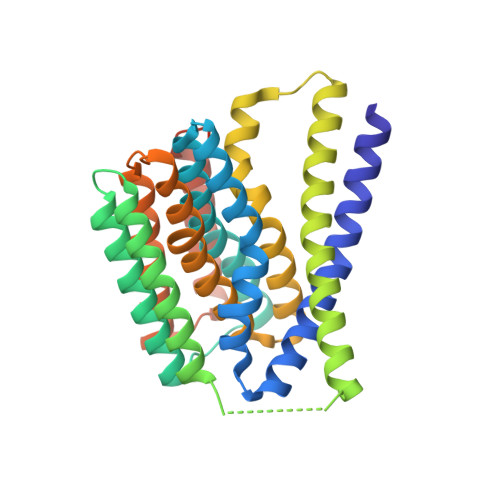
Structural insight into the ion-exchange mechanism of the sodium/calcium exchanger.

Liao, J.Li, H.Zeng, W.Sauer, D.B.Belmares, R.Jiang, Y.

(2012) Science 335: 686-690

  • DOI: https://doi.org/10.1126/science.1215759
  • Primary Citation of Related Structures:  
    3V5S, 3V5U

  • PubMed Abstract: 

    Sodium/calcium (Na(+)/Ca(2+)) exchangers (NCX) are membrane transporters that play an essential role in maintaining the homeostasis of cytosolic Ca(2+) for cell signaling. We demonstrated the Na(+)/Ca(2+)-exchange function of an NCX from Methanococcus jannaschii (NCX_Mj) and report its 1.9 angstrom crystal structure in an outward-facing conformation. Containing 10 transmembrane helices, the two halves of NCX_Mj share a similar structure with opposite orientation. Four ion-binding sites cluster at the center of the protein: one specific for Ca(2+) and three that likely bind Na(+). Two passageways allow for Na(+) and Ca(2+) access to the central ion-binding sites from the extracellular side. Based on the symmetry of NCX_Mj and its ability to catalyze bidirectional ion-exchange reactions, we propose a structure model for the inward-facing NCX_Mj.


  • Organizational Affiliation

    Department of Physiology, University of Texas Southwestern Medical Center, Dallas, TX 75390-9040, USA.


Macromolecules
Find similar proteins by:  (by identity cutoff)  |  3D Structure
Entity ID: 1
MoleculeChains Sequence LengthOrganismDetailsImage
Uncharacterized membrane protein MJ0091320Methanocaldococcus jannaschii DSM 2661Mutation(s): 1 
Gene Names: MJ0091
Membrane Entity: Yes 
UniProt
Find proteins for Q57556 (Methanocaldococcus jannaschii (strain ATCC 43067 / DSM 2661 / JAL-1 / JCM 10045 / NBRC 100440))
Explore Q57556 
Go to UniProtKB:  Q57556
Entity Groups  
Sequence Clusters30% Identity50% Identity70% Identity90% Identity95% Identity100% Identity
UniProt GroupQ57556
Sequence Annotations
Expand
  • Reference Sequence
Small Molecules
Ligands 6 Unique
IDChains Name / Formula / InChI Key2D Diagram3D Interactions
OLC
Query on OLC

Download Ideal Coordinates CCD File 
K [auth A],
L [auth A],
M [auth A],
X [auth A],
Y [auth A]
(2R)-2,3-dihydroxypropyl (9Z)-octadec-9-enoate
C21 H40 O4
RZRNAYUHWVFMIP-GDCKJWNLSA-N
1PE
Query on 1PE

Download Ideal Coordinates CCD File 
Q [auth A]PENTAETHYLENE GLYCOL
C10 H22 O6
JLFNLZLINWHATN-UHFFFAOYSA-N
MYS
Query on MYS

Download Ideal Coordinates CCD File 
N [auth A]
O [auth A]
P [auth A]
R [auth A]
S [auth A]
PENTADECANE
C15 H32
YCOZIPAWZNQLMR-UHFFFAOYSA-N
ACT
Query on ACT

Download Ideal Coordinates CCD File 
G [auth A],
H [auth A],
I [auth A],
J [auth A]
ACETATE ION
C2 H3 O2
QTBSBXVTEAMEQO-UHFFFAOYSA-M
CA
Query on CA

Download Ideal Coordinates CCD File 
C [auth A],
F [auth A]
CALCIUM ION
Ca
BHPQYMZQTOCNFJ-UHFFFAOYSA-N
NA
Query on NA

Download Ideal Coordinates CCD File 
B [auth A],
D [auth A],
E [auth A]
SODIUM ION
Na
FKNQFGJONOIPTF-UHFFFAOYSA-N
Experimental Data & Validation

Experimental Data

  • Method: X-RAY DIFFRACTION
  • Resolution: 1.90 Å
  • R-Value Free:  0.210 (Depositor), 0.210 (DCC) 
  • R-Value Work:  0.185 (Depositor), 0.180 (DCC) 
  • R-Value Observed: 0.187 (Depositor) 
Space Group: P 21 21 21
Unit Cell:
Length ( Å )Angle ( ˚ )
a = 49.49α = 90
b = 72.879β = 90
c = 96.206γ = 90
Software Package:
Software NamePurpose
HKL-3000data collection
SHARPphasing
PHENIXrefinement
HKL-3000data reduction
HKL-3000data scaling

Structure Validation

View Full Validation Report


Ligand Structure Quality Assessment 

Created with Raphaël 2.3.0Worse 01 BetterLigand structure goodness of fit to experimental dataBest fitted OLCClick on this verticalbar to view detailsBest fitted MYSClick on this verticalbar to view details

Entry History 

Deposition Data

Revision History  (Full details and data files)

  • Version 1.0: 2012-02-22
    Type: Initial release
  • Version 1.1: 2024-02-28
    Changes: Data collection, Database references, Derived calculations