3VH8 | pdb_00003vh8

KIR3DL1 in complex with HLA-B*5701


Experimental Data Snapshot

  • Method: X-RAY DIFFRACTION
  • Resolution: 1.80 Å
  • R-Value Free: 
    0.237 (Depositor), 0.240 (DCC) 
  • R-Value Work: 
    0.211 (Depositor), 0.210 (DCC) 
  • R-Value Observed: 
    0.213 (Depositor) 

Starting Models: experimental
View more details

wwPDB Validation   3D Report Full Report


Ligand Structure Quality Assessment 

Created with Raphaël 2.3.0Worse 01 BetterLigand structure goodness of fit to experimental dataBest fitted NAGClick on this verticalbar to view details

This is version 1.4 of the entry. See complete history


Literature

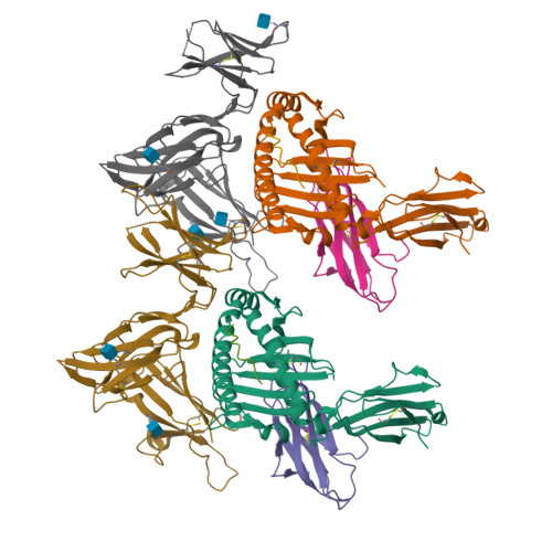
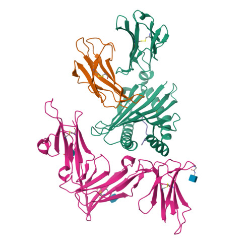
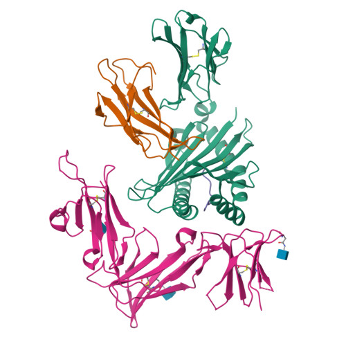
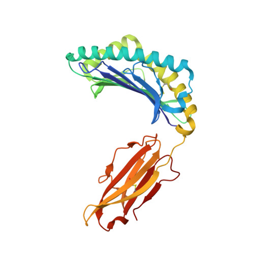
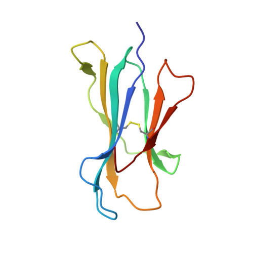
Killer cell immunoglobulin-like receptor 3DL1-mediated recognition of human leukocyte antigen B

Vivian, J.P.Duncan, R.C.Berry, R.O'Connor, G.M.Reid, H.H.Beddoe, T.Gras, S.Saunders, P.M.Olshina, M.A.Widjaja, J.M.L.Harpur, C.M.Lin, J.Maloveste, S.M.Price, D.A.Lafont, B.A.P.McVicar, D.W.Clements, C.S.Brooks, A.G.Rossjohn, J.

(2011) Nature 479: 401-405

  • DOI: https://doi.org/10.1038/nature10517
  • Primary Citation of Related Structures:  
    3VH8

  • PubMed Abstract: 

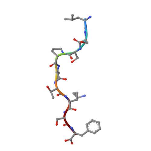
    Members of the killer cell immunoglobulin-like receptor (KIR) family, a large group of polymorphic receptors expressed on natural killer (NK) cells, recognize particular peptide-laden human leukocyte antigen (pHLA) class I molecules and have a pivotal role in innate immune responses. Allelic variation and extensive polymorphism within the three-domain KIR family (KIR3D, domains D0-D1-D2) affects pHLA binding specificity and is linked to the control of viral replication and the treatment outcome of certain haematological malignancies. Here we describe the structure of a human KIR3DL1 receptor bound to HLA-B*5701 complexed with a self-peptide. KIR3DL1 clamped around the carboxy-terminal end of the HLA-B*5701 antigen-binding cleft, resulting in two discontinuous footprints on the pHLA. First, the D0 domain, a distinguishing feature of the KIR3D family, extended towards β2-microglobulin and abutted a region of the HLA molecule with limited polymorphism, thereby acting as an 'innate HLA sensor' domain. Second, whereas the D2-HLA-B*5701 interface exhibited a high degree of complementarity, the D1-pHLA-B*5701 contacts were suboptimal and accommodated a degree of sequence variation both within the peptide and the polymorphic region of the HLA molecule. Although the two-domain KIR (KIR2D) and KIR3DL1 docked similarly onto HLA-C and HLA-B respectively, the corresponding D1-mediated interactions differed markedly, thereby providing insight into the specificity of KIR3DL1 for discrete HLA-A and HLA-B allotypes. Collectively, in association with extensive mutagenesis studies at the KIR3DL1-pHLA-B*5701 interface, we provide a framework for understanding the intricate interplay between peptide variability, KIR3D and HLA polymorphism in determining the specificity requirements of this essential innate interaction that is conserved across primate species.


  • Organizational Affiliation

    Department of Biochemistry and Molecular Biology, School of Biomedical Sciences, Monash University, Clayton, Victoria 3800, Australia.


Macromolecules
Find similar proteins by:  (by identity cutoff)  |  3D Structure
Entity ID: 1
MoleculeChains Sequence LengthOrganismDetailsImage
HLA class I histocompatibility antigen, B-57 alpha chainA,
E [auth D]
275Homo sapiensMutation(s): 0 
Gene Names: HLA-B*5701
UniProt & NIH Common Fund Data Resources
Find proteins for P01889 (Homo sapiens)
Explore P01889 
Go to UniProtKB:  P01889
PHAROS:  P01889
GTEx:  ENSG00000234745 
Entity Groups  
Sequence Clusters30% Identity50% Identity70% Identity90% Identity95% Identity100% Identity
UniProt GroupP01889
Sequence Annotations
Expand
  • Reference Sequence
Find similar proteins by:  (by identity cutoff)  |  3D Structure
Entity ID: 2
MoleculeChains Sequence LengthOrganismDetailsImage
Beta-2-microglobulinB,
F [auth E]
99Homo sapiensMutation(s): 0 
Gene Names: B2MCDABP0092HDCMA22P
UniProt & NIH Common Fund Data Resources
Find proteins for P61769 (Homo sapiens)
Explore P61769 
Go to UniProtKB:  P61769
PHAROS:  P61769
GTEx:  ENSG00000166710 
Entity Groups  
Sequence Clusters30% Identity50% Identity70% Identity90% Identity95% Identity100% Identity
UniProt GroupP61769
Sequence Annotations
Expand
  • Reference Sequence

Find similar proteins by:  Sequence   |   3D Structure  

Entity ID: 3
MoleculeChains Sequence LengthOrganismDetailsImage
peptide of Ig kappa chain C regionC,
G [auth F]
9Homo sapiensMutation(s): 0 
UniProt & NIH Common Fund Data Resources
Find proteins for P01834 (Homo sapiens)
Explore P01834 
Go to UniProtKB:  P01834
PHAROS:  P01834
Entity Groups  
UniProt GroupP01834
Sequence Annotations
Expand
  • Reference Sequence
Find similar proteins by:  (by identity cutoff)  |  3D Structure
Entity ID: 4
MoleculeChains Sequence LengthOrganismDetailsImage
Killer cell immunoglobulin-like receptor 3DL1D [auth G],
H
316Homo sapiensMutation(s): 0 
Gene Names: KIR3DL1*001
UniProt & NIH Common Fund Data Resources
Find proteins for P43629 (Homo sapiens)
Explore P43629 
Go to UniProtKB:  P43629
PHAROS:  P43629
GTEx:  ENSG00000167633 
Entity Groups  
Sequence Clusters30% Identity50% Identity70% Identity90% Identity95% Identity100% Identity
UniProt GroupP43629
Glycosylation
Glycosylation Sites: 3Go to GlyGen: P43629-1
Sequence Annotations
Expand
  • Reference Sequence
Experimental Data & Validation

Experimental Data

  • Method: X-RAY DIFFRACTION
  • Resolution: 1.80 Å
  • R-Value Free:  0.237 (Depositor), 0.240 (DCC) 
  • R-Value Work:  0.211 (Depositor), 0.210 (DCC) 
  • R-Value Observed: 0.213 (Depositor) 
Space Group: P 1
Unit Cell:
Length ( Å )Angle ( ˚ )
a = 66.248α = 100.56
b = 66.374β = 92.83
c = 93.361γ = 101.56
Software Package:
Software NamePurpose
Blu-Icedata collection
PHASERphasing
REFMACrefinement
MOSFLMdata reduction
SCALAdata scaling

Structure Validation

View Full Validation Report


Ligand Structure Quality Assessment 

Created with Raphaël 2.3.0Worse 01 BetterLigand structure goodness of fit to experimental dataBest fitted NAGClick on this verticalbar to view details

Entry History 

Deposition Data

Revision History  (Full details and data files)

  • Version 1.0: 2011-10-26
    Type: Initial release
  • Version 1.1: 2011-12-07
    Changes: Database references
  • Version 1.2: 2020-07-29
    Type: Remediation
    Reason: Carbohydrate remediation
    Changes: Data collection, Database references, Derived calculations, Structure summary
  • Version 1.3: 2023-11-08
    Changes: Data collection, Database references, Refinement description, Structure summary
  • Version 1.4: 2024-11-20
    Changes: Structure summary