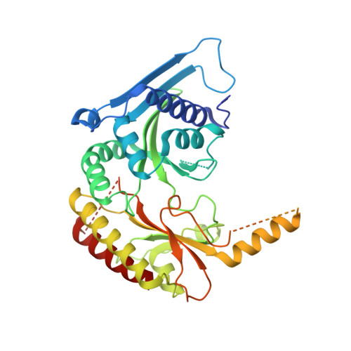
3X04

Crystal structure of PIP4KIIBETA complex with GMPPNP


Experimental Data Snapshot

  • Method: X-RAY DIFFRACTION
  • Resolution: 2.60 Å
  • R-Value Free: 0.277 
  • R-Value Work: 0.229 
  • R-Value Observed: 0.231 

Starting Model: experimental
View more details

wwPDB Validation   3D Report Full Report


Ligand Structure Quality Assessment 


This is version 1.2 of the entry. See complete history


Literature

The Lipid Kinase PI5P4K beta Is an Intracellular GTP Sensor for Metabolism and Tumorigenesis

Sumita, K.Lo, Y.H.Takeuchi, K.Senda, M.Kofuji, S.Ikeda, Y.Terakawa, J.Sasaki, M.Yoshino, H.Majd, N.Zheng, Y.Kahoud, E.R.Yokota, T.Emerling, B.M.Asara, J.M.Ishida, T.Locasale, J.W.Daikoku, T.Anastasiou, D.Senda, T.Sasaki, A.T.

(2016) Mol Cell 61: 187-198

  • DOI: https://doi.org/10.1016/j.molcel.2015.12.011

  • PubMed Abstract: 

    While cellular GTP concentration dramatically changes in response to an organism's cellular status, whether it serves as a metabolic cue for biological signaling remains elusive due to the lack of molecular identification of GTP sensors. Here we report that PI5P4Kβ, a phosphoinositide kinase that regulates PI(5)P levels, detects GTP concentration and converts them into lipid second messenger signaling. Biochemical analyses show that PI5P4Kβ preferentially utilizes GTP, rather than ATP, for PI(5)P phosphorylation, and its activity reflects changes in direct proportion to the physiological GTP concentration. Structural and biological analyses reveal that the GTP-sensing activity of PI5P4Kβ is critical for metabolic adaptation and tumorigenesis. These results demonstrate that PI5P4Kβ is the missing GTP sensor and that GTP concentration functions as a metabolic cue via PI5P4Kβ. The critical role of the GTP-sensing activity of PI5P4Kβ in cancer signifies this lipid kinase as a cancer therapeutic target.


  • Organizational Affiliation

    Division of Hematology and Oncology, Department of Internal Medicine, University of Cincinnati College of Medicine, Cincinnati, OH 45267, USA.


Macromolecules
Find similar proteins by:  (by identity cutoff)  |  3D Structure
Entity ID: 1
MoleculeChains Sequence LengthOrganismDetailsImage
Phosphatidylinositol 5-phosphate 4-kinase type-2 beta
A, B
393Homo sapiensMutation(s): 0 
Gene Names: PIP4K2B
EC: 2.7.1.149
UniProt & NIH Common Fund Data Resources
Find proteins for P78356 (Homo sapiens)
Explore P78356 
Go to UniProtKB:  P78356
PHAROS:  P78356
GTEx:  ENSG00000276293 
Entity Groups  
Sequence Clusters30% Identity50% Identity70% Identity90% Identity95% Identity100% Identity
UniProt GroupP78356
Sequence Annotations
Expand
  • Reference Sequence
Experimental Data & Validation

Experimental Data

  • Method: X-RAY DIFFRACTION
  • Resolution: 2.60 Å
  • R-Value Free: 0.277 
  • R-Value Work: 0.229 
  • R-Value Observed: 0.231 
  • Space Group: C 2 2 21
Unit Cell:
Length ( Å )Angle ( ˚ )
a = 109.804α = 90
b = 185.222β = 90
c = 106.449γ = 90
Software Package:
Software NamePurpose
ADSCdata collection
PHASERphasing
PHENIXrefinement
XDSdata reduction
XSCALEdata scaling

Structure Validation

View Full Validation Report


Ligand Structure Quality Assessment 


Entry History 

Revision History  (Full details and data files)

  • Version 1.0: 2015-10-14
    Type: Initial release
  • Version 1.1: 2016-02-03
    Changes: Database references
  • Version 1.2: 2023-11-08
    Changes: Data collection, Database references, Derived calculations, Refinement description