3WOM

Crystal structure of the DAP BII dipeptide complex II


Experimental Data Snapshot

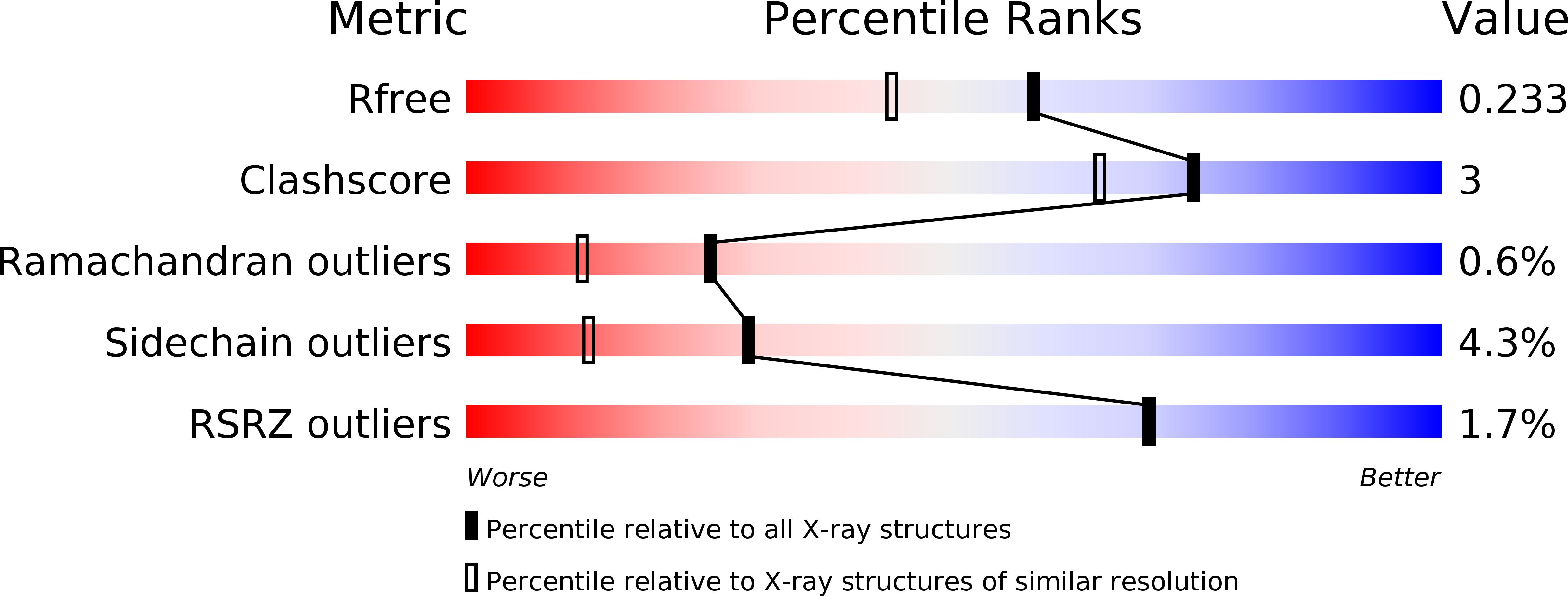
  • Method: X-RAY DIFFRACTION
  • Resolution: 1.86 Å
  • R-Value Free: 0.225 
  • R-Value Work: 0.183 
  • R-Value Observed: 0.185 

Starting Model: experimental
View more details

wwPDB Validation   3D Report Full Report


Ligand Structure Quality Assessment 


This is version 1.1 of the entry. See complete history


Literature

S46 peptidases are the first exopeptidases to be members of clan PA

Sakamoto, Y.Suzuki, Y.Iizuka, I.Tateoka, C.Roppongi, S.Fujimoto, M.Inaka, K.Tanaka, H.Masaki, M.Ohta, K.Okada, H.Nonaka, T.Morikawa, Y.Nakamura, K.T.Ogasawara, W.Tanaka, N.

(2014) Sci Rep 4: 4977-4977

  • DOI: https://doi.org/10.1038/srep04977
  • Primary Citation of Related Structures:  
    3WOI, 3WOJ, 3WOK, 3WOL, 3WOM, 3WON, 3WOO, 3WOP, 3WOQ, 3WOR

  • PubMed Abstract: 

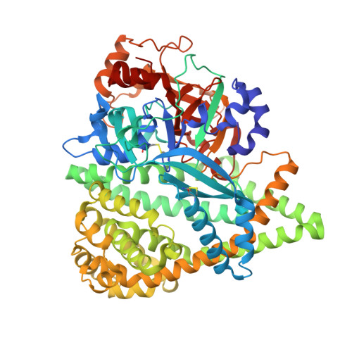
    The dipeptidyl aminopeptidase BII (DAP BII) belongs to a serine peptidase family, S46. The amino acid sequence of the catalytic unit of DAP BII exhibits significant similarity to those of clan PA endopeptidases, such as chymotrypsin. However, the molecular mechanism of the exopeptidase activity of family S46 peptidase is unknown. Here, we report crystal structures of DAP BII. DAP BII contains a peptidase domain including a typical double β-barrel fold and previously unreported α-helical domain. The structures of peptide complexes revealed that the α-helical domain covers the active-site cleft and the side chain of Asn330 in the domain forms hydrogen bonds with the N-terminus of the bound peptide. These observations indicate that the α-helical domain regulates the exopeptidase activity of DAP BII. Because S46 peptidases are not found in mammals, we expect that our study will be useful for the design of specific inhibitors of S46 peptidases from pathogens.


  • Organizational Affiliation

    1] School of Pharmacy, Iwate Medical University, 2-1-1 Nishitokuta, Yahaba, Iwate 028-3694, JAPAN [2].


Macromolecules
Find similar proteins by:  (by identity cutoff)  |  3D Structure
Entity ID: 1
MoleculeChains Sequence LengthOrganismDetailsImage
dipeptidyl aminopeptidase BII
A, B
698Pseudoxanthomonas mexicanaMutation(s): 0 
EC: 3.4.14
UniProt
Find proteins for V5YM14 (Pseudoxanthomonas mexicana)
Explore V5YM14 
Go to UniProtKB:  V5YM14
Entity Groups  
Sequence Clusters30% Identity50% Identity70% Identity90% Identity95% Identity100% Identity
UniProt GroupV5YM14
Sequence Annotations
Expand
  • Reference Sequence
Small Molecules
Ligands 4 Unique
IDChains Name / Formula / InChI Key2D Diagram3D Interactions
TYR
Query on TYR

Download Ideal Coordinates CCD File 
D [auth A],
N [auth B]
TYROSINE
C9 H11 N O3
OUYCCCASQSFEME-QMMMGPOBSA-N
VAL
Query on VAL

Download Ideal Coordinates CCD File 
C [auth A],
M [auth B]
VALINE
C5 H11 N O2
KZSNJWFQEVHDMF-BYPYZUCNSA-N
GOL
Query on GOL

Download Ideal Coordinates CCD File 
E [auth A]
F [auth A]
G [auth A]
H [auth A]
I [auth A]
E [auth A],
F [auth A],
G [auth A],
H [auth A],
I [auth A],
J [auth A],
O [auth B],
P [auth B],
Q [auth B],
R [auth B]
GLYCEROL
C3 H8 O3
PEDCQBHIVMGVHV-UHFFFAOYSA-N
ZN
Query on ZN

Download Ideal Coordinates CCD File 
K [auth A],
L [auth A],
S [auth B],
T [auth B]
ZINC ION
Zn
PTFCDOFLOPIGGS-UHFFFAOYSA-N
Experimental Data & Validation

Experimental Data

  • Method: X-RAY DIFFRACTION
  • Resolution: 1.86 Å
  • R-Value Free: 0.225 
  • R-Value Work: 0.183 
  • R-Value Observed: 0.185 
  • Space Group: P 43 21 2
Unit Cell:
Length ( Å )Angle ( ˚ )
a = 122.22α = 90
b = 122.22β = 90
c = 219.76γ = 90
Software Package:
Software NamePurpose
ADSCdata collection
REFMACrefinement
MOSFLMdata reduction
SCALAdata scaling

Structure Validation

View Full Validation Report


Ligand Structure Quality Assessment 


Entry History 

Deposition Data

Revision History  (Full details and data files)

  • Version 1.0: 2014-09-03
    Type: Initial release
  • Version 1.1: 2023-11-08
    Changes: Data collection, Database references, Derived calculations, Refinement description