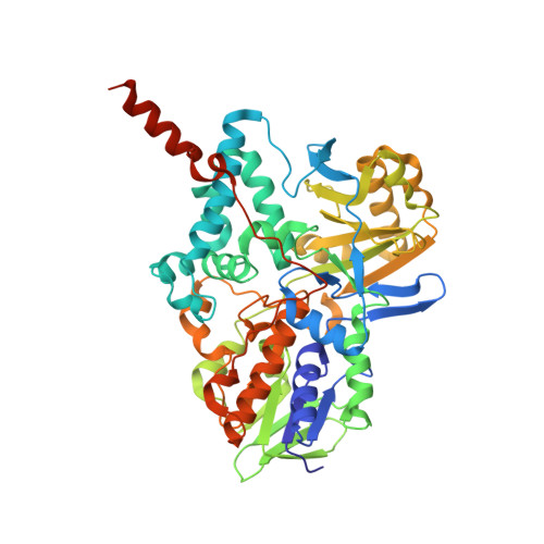
4A7A

Crystal structure of human monoamine oxidase B (MAO B) in complex with rosiglitazone


Experimental Data Snapshot

  • Method: X-RAY DIFFRACTION
  • Resolution: 1.70 Å
  • R-Value Free: 0.214 
  • R-Value Work: 0.186 
  • R-Value Observed: 0.187 

Starting Model: experimental
View more details

wwPDB Validation   3D Report Full Report


Ligand Structure Quality Assessment 


This is version 1.4 of the entry. See complete history


Literature

Molecular Insights Into Human Monoamine Oxidase B Inhibition by the Glitazone Anti-Diabetes Drugs

Binda, C.Aldeco, M.Geldenhuys, W.J.Tortorici, M.Mattevi, A.Edmondson, D.E.

(2012) ACS Med Chem Lett 3: 39-42

  • DOI: https://doi.org/10.1021/ml200196p
  • Primary Citation of Related Structures:  
    4A79, 4A7A

  • PubMed Abstract: 

    The widely employed anti-diabetic drug pioglitazone (Actos) is shown to be a specific and reversible inhibitor of human monoamine oxidase B (MAO B). The crystal structure of the enzyme-inhibitor complex shows the R-enantiomer is bound with the thiazolidinedione ring near the flavin. The molecule occupies both substrate and entrance cavities of the active site establishing non-covalent interactions with the surrounding amino acids. These binding properties differentiate pioglitazone from the clinically used MAO inhibitors, which act through covalent inhibition mechanisms and do not exhibit a high degree of MAO A versus B selectivity. Rosiglitazone (Avandia) and troglitazone, other members of the glitazone class, are less selective in that they are weaker inhibitors of both MAO A and MAO B These results suggest that pioglitazone may have utility as a "re-purposed" neuro-protectant drug in retarding the progression of disease in Parkinson's patients. They also provide new insights for the development of reversible isoenzyme-specific MAO inhibitors.


  • Organizational Affiliation

    Department of Genetics and Microbiology, University of Pavia, 27100 Pavia, Italy.


Macromolecules
Find similar proteins by:  (by identity cutoff)  |  3D Structure
Entity ID: 1
MoleculeChains Sequence LengthOrganismDetailsImage
AMINE OXIDASE [FLAVIN-CONTAINING] B
A, B
520Homo sapiensMutation(s): 0 
EC: 1.4.3.4 (PDB Primary Data), 1.4.3.21 (UniProt)
Membrane Entity: Yes 
UniProt & NIH Common Fund Data Resources
Find proteins for P27338 (Homo sapiens)
Explore P27338 
Go to UniProtKB:  P27338
PHAROS:  P27338
GTEx:  ENSG00000069535 
Entity Groups  
Sequence Clusters30% Identity50% Identity70% Identity90% Identity95% Identity100% Identity
UniProt GroupP27338
Sequence Annotations
Expand
  • Reference Sequence
Experimental Data & Validation

Experimental Data

  • Method: X-RAY DIFFRACTION
  • Resolution: 1.70 Å
  • R-Value Free: 0.214 
  • R-Value Work: 0.186 
  • R-Value Observed: 0.187 
  • Space Group: C 2 2 2
Unit Cell:
Length ( Å )Angle ( ˚ )
a = 131.47α = 90
b = 223.22β = 90
c = 86.53γ = 90
Software Package:
Software NamePurpose
REFMACrefinement
MOSFLMdata reduction
SCALAdata scaling
MOLREPphasing

Structure Validation

View Full Validation Report


Ligand Structure Quality Assessment 


Entry History 

Deposition Data

Revision History  (Full details and data files)

  • Version 1.0: 2012-04-25
    Type: Initial release
  • Version 1.1: 2012-05-30
    Changes: Other
  • Version 1.2: 2012-07-04
    Changes: Other
  • Version 1.3: 2023-12-20
    Changes: Data collection, Database references, Derived calculations, Other, Refinement description
  • Version 1.4: 2024-11-20
    Changes: Structure summary