4AJ9 | pdb_00004aj9

Catalase 3 from Neurospora crassa


Experimental Data Snapshot

  • Method: X-RAY DIFFRACTION
  • Resolution: 1.85 Å
  • R-Value Free: 
    0.218 (Depositor), 0.210 (DCC) 
  • R-Value Work: 
    0.177 (Depositor), 0.170 (DCC) 
  • R-Value Observed: 
    0.179 (Depositor) 

Starting Model: experimental
View more details

wwPDB Validation   3D Report Full Report


Ligand Structure Quality Assessment 

Created with Raphaël 2.3.0Worse 01 BetterLigand structure goodness of fit to experimental dataBest fitted HEMClick on this verticalbar to view details

This is version 1.2 of the entry. See complete history


Literature

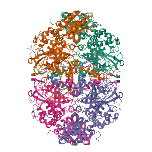
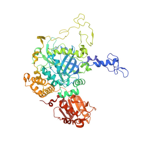
X-ray driven reduction of Cpd I of Catalase-3 from N. crassa reveals differential sensitivity of active sites and formation of ferrous state.

Zarate-Romero, A.Stojanoff, V.Cohen, A.E.Hansberg, W.Rudino-Pinera, E.

(2019) Arch Biochem Biophys 666: 107-115

  • DOI: https://doi.org/10.1016/j.abb.2019.03.020
  • Primary Citation of Related Structures:  
    4AJ9, 6NSW, 6NSY, 6NSZ, 6NT0, 6NT1

  • PubMed Abstract: 

    Catalases are biotechnologically relevant enzymes because of their applications in food technology, bioremediation, and biomedicine. The dismutation of hydrogen peroxide occurs in two steps; in the first one, the enzyme forms an oxidized compound I (Cpd I) and in the second one, the enzyme is reduced to the ferric state. In this research work, we analyzed the reduction of Cpd I by X-ray radiation damage during diffraction experiments in crystals of CAT-3, a Large-Size Subunit Catalase (LSC) from Neurospora crassa. A Multi-Crystal Data collection Strategy was applied in order to obtain the Cpd I structure at a resolution of 2.2 Å; this intermediate was highly sensitive to X-ray and was easily reduced at very low deposited radiation dose, causing breakage of the Fe=O bond. The comparison of the structures showed reduced intermediates and also evidenced the differential sensitivity per monomer. The resting ferric state was reduced to the ferrous state, an intermediate without a previous report in LSC. The chemically obtained Cpd I and the X-ray reduced intermediates were identified by UV-visible microspectrometry coupled to data collection. The differential sensitivity and the formation of a ferrous state are discussed, emphasizing the importance of the correct interpretation in the oxidation state of the iron heme.


  • Organizational Affiliation

    Cátedras CONACyT, Departamento de Bionanotecnología, Centro de Nanociencias y Nanotecnología, Universidad Nacional Autónoma de México, Ensenada, B. C., Mexico.


Macromolecules
Find similar proteins by:  (by identity cutoff)  |  3D Structure
Entity ID: 1
MoleculeChains Sequence LengthOrganismDetailsImage
CATALASE-3
A, B, C, D
682Neurospora crassaMutation(s): 0 
EC: 1.11.1.6
UniProt
Find proteins for Q9C169 (Neurospora crassa (strain ATCC 24698 / 74-OR23-1A / CBS 708.71 / DSM 1257 / FGSC 987))
Explore Q9C169 
Go to UniProtKB:  Q9C169
Entity Groups  
Sequence Clusters30% Identity50% Identity70% Identity90% Identity95% Identity100% Identity
UniProt GroupQ9C169
Sequence Annotations
Expand
  • Reference Sequence
Small Molecules
Ligands 3 Unique
IDChains Name / Formula / InChI Key2D Diagram3D Interactions
HEM
Query on HEM

Download Ideal Coordinates CCD File 
E [auth A],
GA [auth D],
O [auth B],
Y [auth C]
PROTOPORPHYRIN IX CONTAINING FE
C34 H32 Fe N4 O4
KABFMIBPWCXCRK-RGGAHWMASA-L
1PE
Query on 1PE

Download Ideal Coordinates CCD File 
BA [auth C]
CA [auth C]
DA [auth C]
EA [auth C]
FA [auth C]
PENTAETHYLENE GLYCOL
C10 H22 O6
JLFNLZLINWHATN-UHFFFAOYSA-N
ACT
Query on ACT

Download Ideal Coordinates CCD File 
AA [auth C]
F [auth A]
G [auth A]
HA [auth D]
IA [auth D]
ACETATE ION
C2 H3 O2
QTBSBXVTEAMEQO-UHFFFAOYSA-M
Experimental Data & Validation

Experimental Data

  • Method: X-RAY DIFFRACTION
  • Resolution: 1.85 Å
  • R-Value Free:  0.218 (Depositor), 0.210 (DCC) 
  • R-Value Work:  0.177 (Depositor), 0.170 (DCC) 
  • R-Value Observed: 0.179 (Depositor) 
Space Group: P 21 21 21
Unit Cell:
Length ( Å )Angle ( ˚ )
a = 130.75α = 90
b = 154.19β = 90
c = 160.3γ = 90
Software Package:
Software NamePurpose
PHENIXrefinement
XDSdata reduction
XSCALEdata scaling
PHASERphasing

Structure Validation

View Full Validation Report


Ligand Structure Quality Assessment 

Created with Raphaël 2.3.0Worse 01 BetterLigand structure goodness of fit to experimental dataBest fitted HEMClick on this verticalbar to view details

Entry History 

Deposition Data

Revision History  (Full details and data files)

  • Version 1.0: 2013-03-06
    Type: Initial release
  • Version 1.1: 2019-04-24
    Changes: Data collection, Database references, Other
  • Version 1.2: 2023-12-20
    Changes: Data collection, Database references, Derived calculations, Other, Refinement description