4CBK | pdb_00004cbk

The c-ring ion binding site of the ATP synthase from Bacillus pseudofirmus OF4 is adapted to alkaliphilic cell physiology


Experimental Data Snapshot

  • Method: X-RAY DIFFRACTION
  • Resolution: 2.42 Å
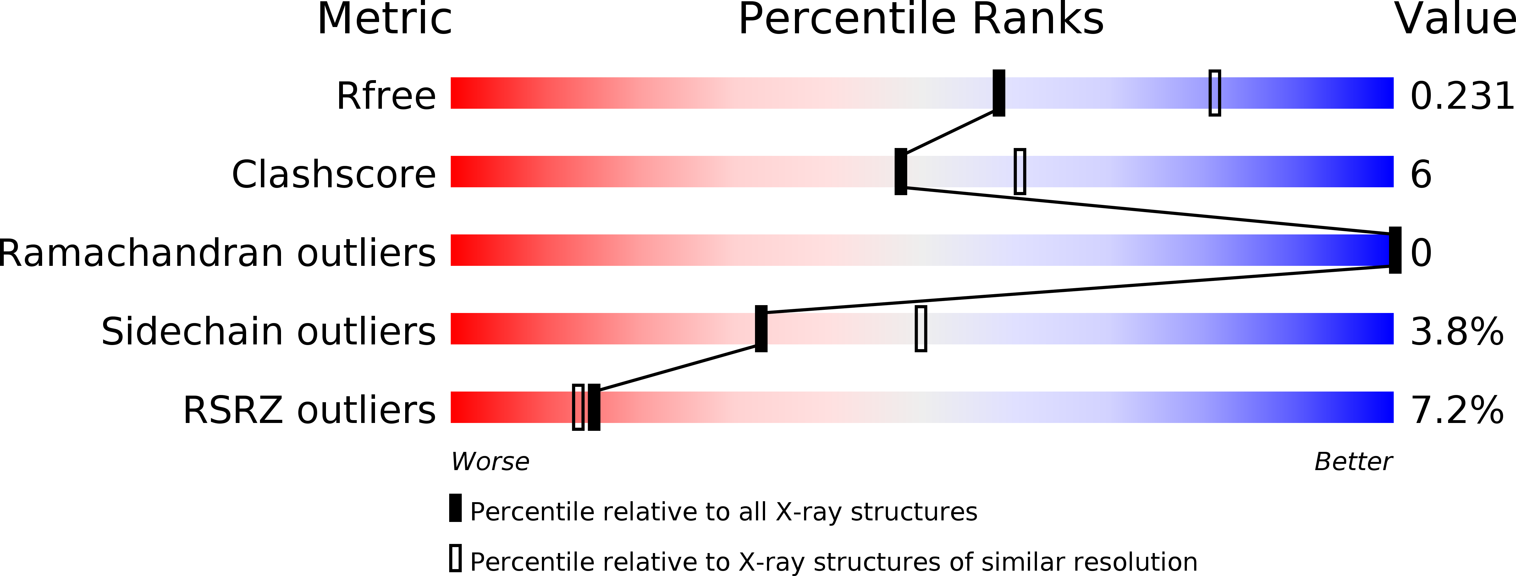
  • R-Value Free: 
    0.228 (Depositor), 0.230 (DCC) 
  • R-Value Work: 
    0.195 (Depositor), 0.200 (DCC) 
  • R-Value Observed: 
    0.197 (Depositor) 

Starting Model: experimental
View more details

wwPDB Validation   3D Report Full Report


Ligand Structure Quality Assessment 

Created with Raphaël 2.3.0Worse 01 BetterLigand structure goodness of fit to experimental dataBest fitted LMTClick on this verticalbar to view detailsBest fitted DPVClick on this verticalbar to view details

This is version 1.3 of the entry. See complete history


Literature

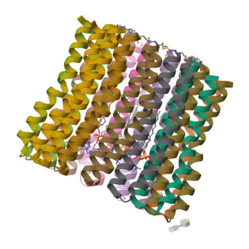
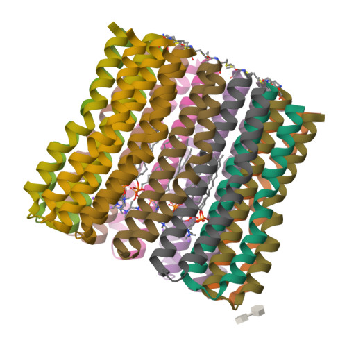
The C-Ring Ion-Binding Site of the ATP Synthase from Bacillus Pseudofirmus of4 is Adapted to Alkaliphilic Lifestyle.

Preiss, L.Langer, J.D.Hicks, D.B.Liu, J.Yildiz, O.Krulwich, T.A.Meier, T.

(2014) Mol Microbiol 92: 973

  • DOI: https://doi.org/10.1111/mmi.12605
  • Primary Citation of Related Structures:  
    4CBJ, 4CBK

  • PubMed Abstract: 

    In the c-ring rotor of ATP synthases ions are shuttled across the membrane during ATP synthesis by a unique rotary mechanism. We investigated characteristics of the c-ring from the alkaliphile Bacillus pseudofirmus OF4 with respect to evolutionary adaptations to operate with protons at high environmental pH. The X-ray structures of the wild-type c13 ring at pH 9.0 and a 'neutralophile-like' mutant (P51A) at pH 4.4, at 2.4 and 2.8 Å resolution, respectively, reveal a dependency of the conformation and protonation state of the proton-binding glutamate (E(54) ) on environmental hydrophobicity. Faster labelling kinetics with the inhibitor dicyclohexylcarbodiimide (DCCD) demonstrate a greater flexibility of E(54) in the mutant due to reduced water occupancy within the H(+) binding site. A second 'neutralophile-like' mutant (V21N) shows reduced growth at high pH, which is explained by restricted conformational freedom of the mutant's E(54) carboxylate. The study directly connects subtle structural adaptations of the c-ring ion binding site to in vivo effects of alkaliphile cell physiology.


  • Organizational Affiliation

    Department of Structural Biology, Max Planck Institute of Biophysics, Max-von-Laue-Str. 3, 60438, Frankfurt am Main, Germany.


Macromolecules
Find similar proteins by:  (by identity cutoff)  |  3D Structure
Entity ID: 1
MoleculeChains Sequence LengthOrganismDetailsImage
ATP SYNTHASE SUBUNIT C
A, B, C, D, E
69Alkalihalophilus pseudofirmus OF4Mutation(s): 0 
Membrane Entity: Yes 
UniProt
Find proteins for P22483 (Alkalihalophilus pseudofirmus (strain ATCC BAA-2126 / JCM 17055 / OF4))
Explore P22483 
Go to UniProtKB:  P22483
Entity Groups  
Sequence Clusters30% Identity50% Identity70% Identity90% Identity95% Identity100% Identity
UniProt GroupP22483
Sequence Annotations
Expand
  • Reference Sequence
Small Molecules
Ligands 3 Unique
IDChains Name / Formula / InChI Key2D Diagram3D Interactions
LMT
Query on LMT

Download Ideal Coordinates CCD File 
Q [auth B]DODECYL-BETA-D-MALTOSIDE
C24 H46 O11
NLEBIOOXCVAHBD-QKMCSOCLSA-N
DPV
Query on DPV

Download Ideal Coordinates CCD File 
AA [auth G]
CA [auth H]
EA [auth I]
FA [auth J]
HA [auth K]
dodecyl 2-(trimethylammonio)ethyl phosphate
C17 H38 N O4 P
QBHFVMDLPTZDOI-UHFFFAOYSA-N
NA
Query on NA

Download Ideal Coordinates CCD File 
BA [auth H]
DA [auth I]
GA [auth K]
IA [auth L]
O [auth B]
SODIUM ION
Na
FKNQFGJONOIPTF-UHFFFAOYSA-N
Modified Residues  1 Unique
IDChains TypeFormula2D DiagramParent
FME
Query on FME
A, B, C, D, E
L-PEPTIDE LINKINGC6 H11 N O3 SMET
Experimental Data & Validation

Experimental Data

  • Method: X-RAY DIFFRACTION
  • Resolution: 2.42 Å
  • R-Value Free:  0.228 (Depositor), 0.230 (DCC) 
  • R-Value Work:  0.195 (Depositor), 0.200 (DCC) 
  • R-Value Observed: 0.197 (Depositor) 
Space Group: P 1 21 1
Unit Cell:
Length ( Å )Angle ( ˚ )
a = 73.79α = 90
b = 96.75β = 105.38
c = 118.33γ = 90
Software Package:
Software NamePurpose
PHENIXrefinement
XDSdata reduction
XSCALEdata scaling
PHASERphasing

Structure Validation

View Full Validation Report


Ligand Structure Quality Assessment 

Created with Raphaël 2.3.0Worse 01 BetterLigand structure goodness of fit to experimental dataBest fitted LMTClick on this verticalbar to view detailsBest fitted DPVClick on this verticalbar to view details

Entry History 

Deposition Data

Revision History  (Full details and data files)

  • Version 1.0: 2014-04-23
    Type: Initial release
  • Version 1.1: 2014-06-11
    Changes: Database references
  • Version 1.2: 2023-12-20
    Changes: Data collection, Database references, Derived calculations, Other, Refinement description
  • Version 1.3: 2024-10-09
    Changes: Structure summary