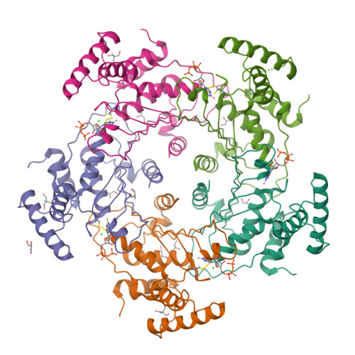
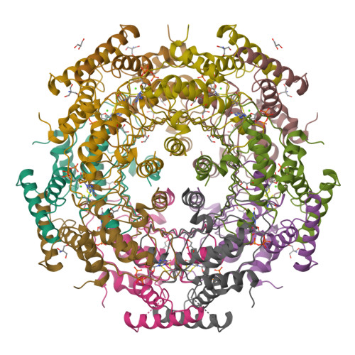
Crystal structure of GTP cyclohydrolase I from Yersinia pestis complexed with GTP
Maltseva, N., Kim, Y., Kwon, K., Anderson, W.F., Joachimiak, A., CSGIDTo be published.
Experimental Data Snapshot
Starting Model: experimental
View more details
Entity ID: 1 | |||||
|---|---|---|---|---|---|
| Molecule | Chains | Sequence Length | Organism | Details | Image |
| GTP cyclohydrolase 1 | 223 | Yersinia pestis CO92 | Mutation(s): 0 Gene Names: folE, YPTB1520 EC: 3.5.4.16 | 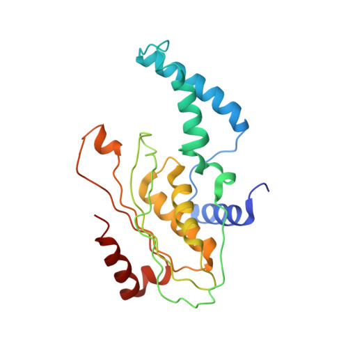 | |
UniProt | |||||
Find proteins for Q8ZG15 (Yersinia pestis) Explore Q8ZG15 Go to UniProtKB: Q8ZG15 | |||||
Entity Groups | |||||
| Sequence Clusters | 30% Identity50% Identity70% Identity90% Identity95% Identity100% Identity | ||||
| UniProt Group | Q8ZG15 | ||||
Sequence AnnotationsExpand | |||||
| |||||
| Ligands 5 Unique | |||||
|---|---|---|---|---|---|
| ID | Chains | Name / Formula / InChI Key | 2D Diagram | 3D Interactions | |
| GTP Query on GTP | F [auth A], K [auth B], N [auth C], T [auth D], X [auth E] | GUANOSINE-5'-TRIPHOSPHATE C10 H16 N5 O14 P3 XKMLYUALXHKNFT-UUOKFMHZSA-N |  | ||
| TRS Query on TRS | J [auth B], O [auth C], U [auth D] | 2-AMINO-2-HYDROXYMETHYL-PROPANE-1,3-DIOL C4 H12 N O3 LENZDBCJOHFCAS-UHFFFAOYSA-O |  | ||
| PEG Query on PEG | I [auth B] | DI(HYDROXYETHYL)ETHER C4 H10 O3 MTHSVFCYNBDYFN-UHFFFAOYSA-N |  | ||
| GOL Query on GOL | G [auth A] H [auth B] M [auth C] Q [auth C] R [auth C] | GLYCEROL C3 H8 O3 PEDCQBHIVMGVHV-UHFFFAOYSA-N |  | ||
| CA Query on CA | L [auth B], P [auth C], V [auth D] | CALCIUM ION Ca BHPQYMZQTOCNFJ-UHFFFAOYSA-N |  | ||
| Length ( Å ) | Angle ( ˚ ) |
|---|---|
| a = 174.047 | α = 90 |
| b = 104.912 | β = 96.89 |
| c = 70.07 | γ = 90 |
| Software Name | Purpose |
|---|---|
| HKL-3000 | data collection |
| HKL-3000 | phasing |
| MOLREP | phasing |
| PHENIX | refinement |
| HKL-3000 | data reduction |
| HKL-3000 | data scaling |