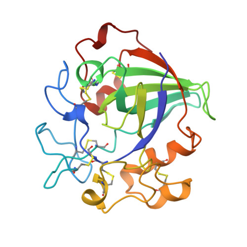
4ENG

STRUCTURE OF ENDOGLUCANASE V CELLOHEXAOSE COMPLEX


Experimental Data Snapshot

  • Method: X-RAY DIFFRACTION
  • Resolution: 1.90 Å
  • R-Value Work: 0.157 

Starting Model: experimental
View more details

wwPDB Validation   3D Report Full Report


This is version 2.3 of the entry. See complete history


Literature

Structure determination and refinement of the Humicola insolens endoglucanase V at 1.5 A resolution.

Davies, G.J.Dodson, G.Moore, M.H.Tolley, S.P.Dauter, Z.Wilson, K.S.Rasmussen, G.Schulein, M.

(1996) Acta Crystallogr D Biol Crystallogr 52: 7-17

  • DOI: https://doi.org/10.1107/S0907444995009280
  • Primary Citation of Related Structures:  
    3ENG, 4ENG

  • PubMed Abstract: 

    The structure of the catalytic core of the endoglucanase V (EGV) from Humicola insolens has been determined by the method of multiple isomorphous replacement at 1.5 A resolution. The final model, refined with X-PLOR and PROLSQ, has a crystallographic R factor of 0.163 (R(free) = 0.240) with deviations from stereochemical target values of 0.012 A and 0.037 degrees for bonds and angles, respectively. The model was further refined with SHELXL, including anisotropic modelling of the protein-atom temperature factors, to give a final model with an R factor of 0.105 and an R(free) of 0.154. The initial isomorphous replacement electron-density map was poor and uninterpretable but was improved by the use of synchrotron data collected at a wavelength chosen so as to optimize the f" contribution of the anomalous scattering from the heavy atoms. The structure of H. insolens EGV consists of a six-stranded beta-barrel domain, similar to that found in a family of plant defence proteins, linked by a number of disulfide-bonded loop regions. A long open groove runs across the surface of the enzyme either side of which lie the catalytic aspartate residues. The 9 A separation of the catalytic carboxylate groups is consistent with the observation that EGV catalyzes the hydrolysis of the cellulose, beta(1-->4) links with inversion of configuration at the anomeric C1 atom. This structure is the first representative from the glycosyl hydrolase family 45.


  • Organizational Affiliation

    Department of Chemistry, University of York, Heslington, England.


Macromolecules
Find similar proteins by:  (by identity cutoff)  |  3D Structure
Entity ID: 1
MoleculeChains Sequence LengthOrganismDetailsImage
ENDOGLUCANASE V CELLOHEXAOSE COMPLEX210Mycothermus thermophilusMutation(s): 1 
EC: 3.2.1.4
UniProt
Find proteins for P43316 (Humicola insolens)
Explore P43316 
Go to UniProtKB:  P43316
Entity Groups  
Sequence Clusters30% Identity50% Identity70% Identity90% Identity95% Identity100% Identity
UniProt GroupP43316
Sequence Annotations
Expand
  • Reference Sequence
Oligosaccharides

Help

Entity ID: 2
MoleculeChains Length2D Diagram Glycosylation3D Interactions
beta-D-glucopyranose-(1-4)-beta-D-glucopyranose-(1-4)-alpha-D-glucopyranose
B, C
3N/A
Glycosylation Resources
GlyTouCan:  G03861VK
GlyCosmos:  G03861VK
GlyGen:  G03861VK
Biologically Interesting Molecules (External Reference) 1 Unique
Experimental Data & Validation

Experimental Data

  • Method: X-RAY DIFFRACTION
  • Resolution: 1.90 Å
  • R-Value Work: 0.157 
  • Space Group: P 1 21 1
Unit Cell:
Length ( Å )Angle ( ˚ )
a = 41.93α = 90
b = 51.98β = 97.73
c = 42.01γ = 90
Software Package:
Software NamePurpose
DENZOdata reduction
ROTAVATAdata reduction
AMoREphasing
REFMACrefinement
CCP4data scaling

Structure Validation

View Full Validation Report


Entry History 

Deposition Data

Revision History  (Full details and data files)

  • Version 1.0: 1997-06-16
    Type: Initial release
  • Version 1.1: 2008-03-03
    Changes: Version format compliance
  • Version 1.2: 2011-07-13
    Changes: Non-polymer description, Version format compliance
  • Version 1.3: 2018-05-02
    Changes: Data collection
  • Version 2.0: 2020-07-29
    Type: Remediation
    Reason: Carbohydrate remediation
    Changes: Advisory, Atomic model, Data collection, Derived calculations, Non-polymer description, Structure summary
  • Version 2.1: 2021-11-03
    Changes: Database references, Structure summary
  • Version 2.2: 2023-08-09
    Changes: Refinement description
  • Version 2.3: 2024-11-13
    Changes: Data collection, Structure summary