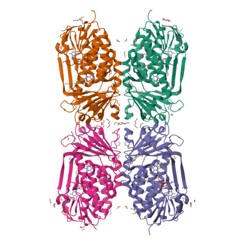
Crystal structure of 2-methyl-3-hydroxypyridine-5-carboxylic acid oxygenase
Kobayashi, J., Yoshida, H., Mikami, B., Hayashi, H., Kamitori, S., Yagi, T.To be published.
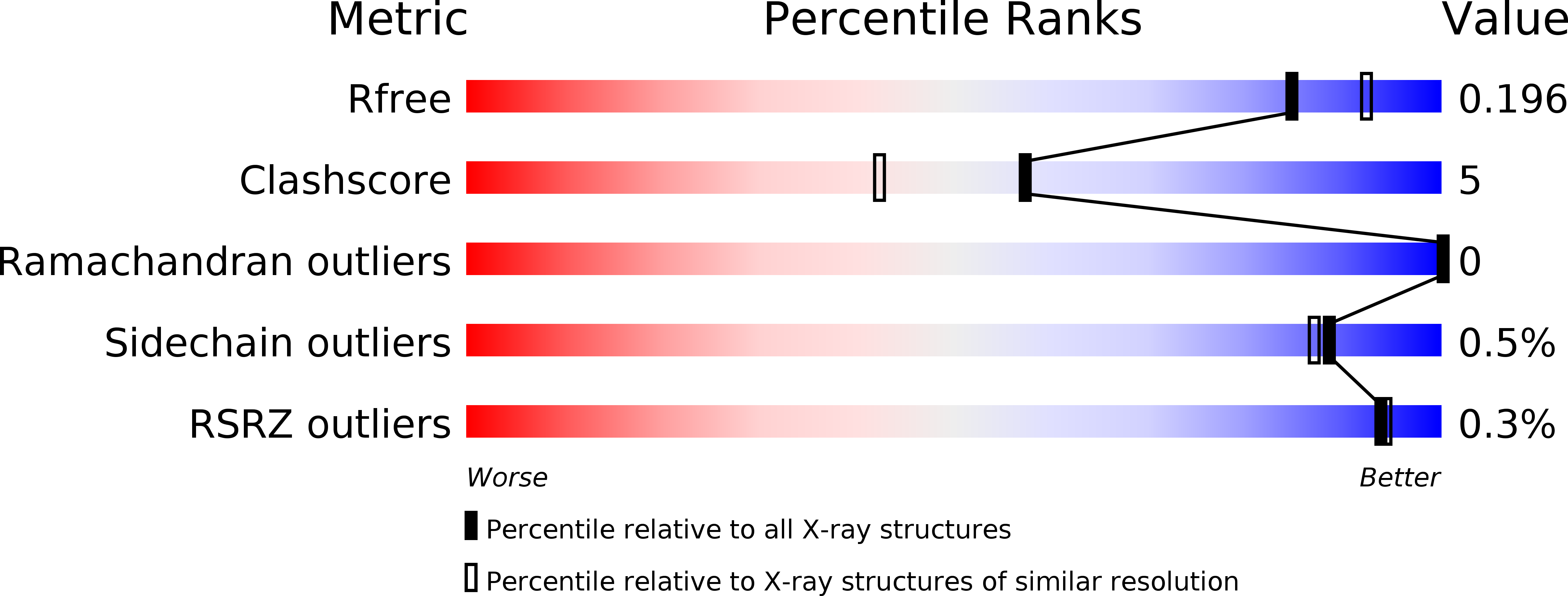
Experimental Data Snapshot
Starting Model: experimental
View more details
Entity ID: 1 | |||||
|---|---|---|---|---|---|
| Molecule | Chains | Sequence Length | Organism | Details | Image |
| 2-methyl-3-hydroxypyridine-5-carboxylic acid oxygenase | 379 | Mesorhizobium japonicum MAFF 303099 | Mutation(s): 0 Gene Names: mlr6788 EC: 1.14.12.4 | 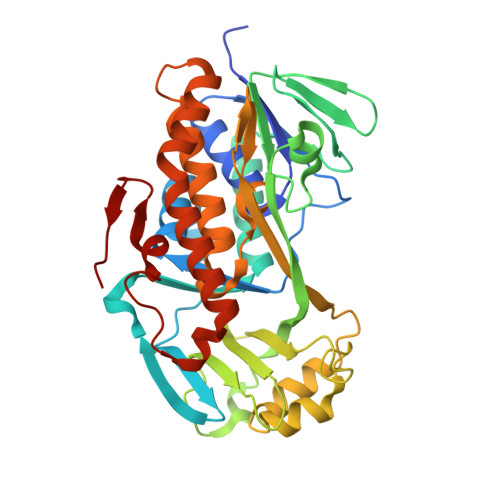 | |
UniProt | |||||
Find proteins for Q988D3 (Mesorhizobium japonicum (strain LMG 29417 / CECT 9101 / MAFF 303099)) Explore Q988D3 Go to UniProtKB: Q988D3 | |||||
Entity Groups | |||||
| Sequence Clusters | 30% Identity50% Identity70% Identity90% Identity95% Identity100% Identity | ||||
| UniProt Group | Q988D3 | ||||
Sequence AnnotationsExpand | |||||
| |||||
| Ligands 7 Unique | |||||
|---|---|---|---|---|---|
| ID | Chains | Name / Formula / InChI Key | 2D Diagram | 3D Interactions | |
| FAD Query on FAD | BB [auth C], E [auth A], X [auth B], ZB [auth D] | FLAVIN-ADENINE DINUCLEOTIDE C27 H33 N9 O15 P2 VWWQXMAJTJZDQX-UYBVJOGSSA-N |  | ||
| PG4 Query on PG4 | AB [auth B], XC [auth D], YB [auth C] | TETRAETHYLENE GLYCOL C8 H18 O5 UWHCKJMYHZGTIT-UHFFFAOYSA-N |  | ||
| PGE Query on PGE | W [auth A], ZA [auth B] | TRIETHYLENE GLYCOL C6 H14 O4 ZIBGPFATKBEMQZ-UHFFFAOYSA-N |  | ||
| PEG Query on PEG | Q [auth A] R [auth A] RA [auth B] S [auth A] SA [auth B] | DI(HYDROXYETHYL)ETHER C4 H10 O3 MTHSVFCYNBDYFN-UHFFFAOYSA-N |  | ||
| SO4 Query on SO4 | AA [auth B] BA [auth B] BC [auth D] CC [auth D] DB [auth C] | SULFATE ION O4 S QAOWNCQODCNURD-UHFFFAOYSA-L |  | ||
| BME Query on BME | AC [auth D], CB [auth C], F [auth A], Y [auth B] | BETA-MERCAPTOETHANOL C2 H6 O S DGVVWUTYPXICAM-UHFFFAOYSA-N |  | ||
| EDO Query on EDO | CA [auth B] DA [auth B] EA [auth B] FA [auth B] FC [auth D] | 1,2-ETHANEDIOL C2 H6 O2 LYCAIKOWRPUZTN-UHFFFAOYSA-N |  | ||
| Length ( Å ) | Angle ( ˚ ) |
|---|---|
| a = 103.838 | α = 90 |
| b = 103.838 | β = 90 |
| c = 452.781 | γ = 120 |
| Software Name | Purpose |
|---|---|
| HKL-2000 | data collection |
| PHENIX | refinement |
| HKL-2000 | data reduction |
| HKL-2000 | data scaling |