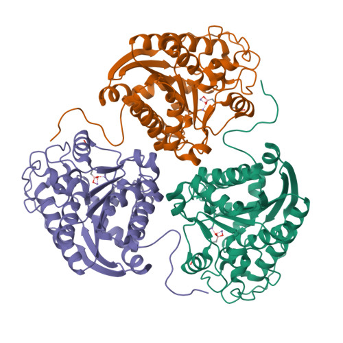
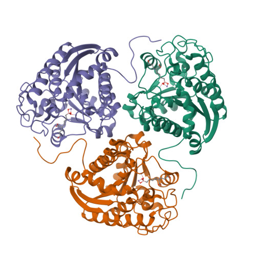
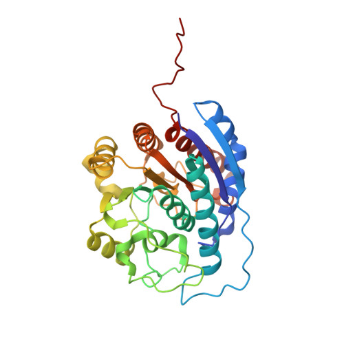
4IE1

Crystal structure of human Arginase-1 complexed with inhibitor 1h


Experimental Data Snapshot

  • Method: X-RAY DIFFRACTION
  • Resolution: 2.00 Å
  • R-Value Free: 0.127 
  • R-Value Work: 0.106 
  • R-Value Observed: 0.108 

Starting Model: experimental
View more details

wwPDB Validation   3D Report Full Report


Ligand Structure Quality Assessment 


This is version 1.4 of the entry. See complete history


Literature

2-Substituted-2-amino-6-boronohexanoic acids as arginase inhibitors.

Golebiowski, A.Paul Beckett, R.Van Zandt, M.Ji, M.K.Whitehouse, D.Ryder, T.R.Jagdmann, E.Andreoli, M.Mazur, A.Padmanilayam, M.Cousido-Siah, A.Mitschler, A.Ruiz, F.X.Podjarny, A.Schroeter, H.

(2013) Bioorg Med Chem Lett 23: 2027-2030

  • DOI: https://doi.org/10.1016/j.bmcl.2013.02.024
  • Primary Citation of Related Structures:  
    4IE1, 4IE2, 4IE3

  • PubMed Abstract: 

    Substitution at the alpha center of the known human arginase inhibitor 2-amino-6-boronohexanoic acid (ABH) is acceptable in the active site pockets of both human arginase I and arginase II. In particular, substituents with a tertiary amine linked via a two carbon chain show improved inhibitory potency for both enzyme isoforms. This potency improvement can be rationalized by X-ray crystallography, which shows a water-mediated contact between the basic nitrogen and the carboxylic acid side chain of Asp200, which is situated at the mouth of the active site pocket of arginase II (Asp181 in arginase I). We believe that this is the first literature report of compounds with improved arginase inhibitory activity, relative to ABH, and represents a promising starting point for further optimization of in vitro potency and the identification of better tool molecules for in vivo investigations of the potential pathophysiological roles of arginases.


  • Organizational Affiliation

    Institutes for Pharmaceutical Discovery, 23 Business Park Drive, Branford, CT, USA. golebiowski.a@gmail.com


Macromolecules
Find similar proteins by:  (by identity cutoff)  |  3D Structure
Entity ID: 1
MoleculeChains Sequence LengthOrganismDetailsImage
Arginase-1
A, B
314Homo sapiensMutation(s): 0 
Gene Names: ARG1
EC: 3.5.3.1
UniProt & NIH Common Fund Data Resources
Find proteins for P05089 (Homo sapiens)
Explore P05089 
Go to UniProtKB:  P05089
PHAROS:  P05089
GTEx:  ENSG00000118520 
Entity Groups  
Sequence Clusters30% Identity50% Identity70% Identity90% Identity95% Identity100% Identity
UniProt GroupP05089
Sequence Annotations
Expand
  • Reference Sequence
Experimental Data & Validation

Experimental Data

  • Method: X-RAY DIFFRACTION
  • Resolution: 2.00 Å
  • R-Value Free: 0.127 
  • R-Value Work: 0.106 
  • R-Value Observed: 0.108 
  • Space Group: P 3
Unit Cell:
Length ( Å )Angle ( ˚ )
a = 90.074α = 90
b = 90.074β = 90
c = 69.272γ = 120
Software Package:
Software NamePurpose
DENZOdata reduction
SCALEPACKdata scaling
AMoREphasing
PHENIXrefinement
PDB_EXTRACTdata extraction
HKL-2000data collection
HKL-2000data reduction
HKL-2000data scaling

Structure Validation

View Full Validation Report


Ligand Structure Quality Assessment 


Entry History 

Revision History  (Full details and data files)

  • Version 1.0: 2013-03-20
    Type: Initial release
  • Version 1.1: 2013-04-03
    Changes: Database references
  • Version 1.2: 2013-12-18
    Changes: Structure summary
  • Version 1.3: 2017-11-15
    Changes: Refinement description
  • Version 1.4: 2023-09-20
    Changes: Data collection, Database references, Derived calculations, Refinement description