4IZ5 | pdb_00004iz5

Structure of the complex between ERK2 phosphomimetic mutant and PEA-15


Experimental Data Snapshot

  • Method: X-RAY DIFFRACTION
  • Resolution: 3.19 Å
  • R-Value Free: 
    0.292 (Depositor), 0.290 (DCC) 
  • R-Value Work: 
    0.243 (Depositor), 0.240 (DCC) 
  • R-Value Observed: 
    0.245 (Depositor) 

wwPDB Validation   3D Report Full Report


Ligand Structure Quality Assessment 

Created with Raphaël 2.3.0Worse 01 BetterLigand structure goodness of fit to experimental dataBest fitted ADPClick on this verticalbar to view details

This is version 1.2 of the entry. See complete history


Literature

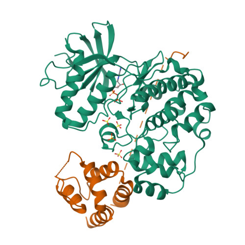
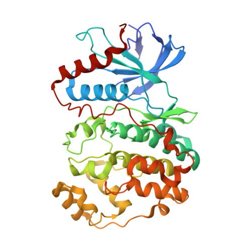
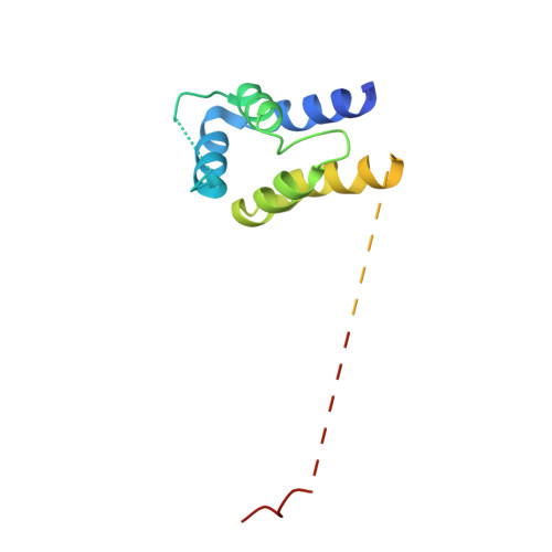
Structure of ERK2 bound to PEA-15 reveals a mechanism for rapid release of activated MAPK.

Mace, P.D.Wallez, Y.Egger, M.F.Dobaczewska, M.K.Robinson, H.Pasquale, E.B.Riedl, S.J.

(2013) Nat Commun 4: 1681-1681

  • DOI: https://doi.org/10.1038/ncomms2687
  • Primary Citation of Related Structures:  
    4IZ5, 4IZ7, 4IZA

  • PubMed Abstract: 

    ERK1/2 kinases are the principal effectors of a central signalling cascade that converts extracellular stimuli into cell proliferation and migration responses and, when deregulated, can promote cell oncogenic transformation. The scaffolding protein PEA-15 is a death effector domain protein that directly interacts with ERK1/2 and affects ERK1/2 subcellular localization and phosphorylation. Here, to understand this ERK1/2 signalling complex, we have solved the crystal structures of PEA-15 bound to three different ERK2 phospho-conformers. The structures reveal that PEA-15 uses a bipartite binding mode, occupying two key docking sites of ERK2. Remarkably, PEA-15 can efficiently bind the ERK2 activation loop in the critical Thr-X-Tyr region in different phosphorylation states. PEA-15 binding triggers an extended allosteric conduit in dually phosphorylated ERK2, disrupting key features of active ERK2. At the same time PEA-15 binding protects ERK2 from dephosphorylation, thus setting the stage for immediate ERK activity upon its release from the PEA-15 inhibitory complex.


  • Organizational Affiliation

    Program in Apoptosis and Cell Death Research, Cancer Center, Sanford-Burnham Medical Research Institute, 10901 North Torrey Pines Road, La Jolla, California 92037, USA.


Macromolecules
Find similar proteins by:  (by identity cutoff)  |  3D Structure
Entity ID: 1
MoleculeChains Sequence LengthOrganismDetailsImage
Mitogen-activated protein kinase 1
A, B, C, D
356Homo sapiensMutation(s): 1 
Gene Names: MAPK1ERK2PRKM1PRKM2
EC: 2.7.11.24
UniProt & NIH Common Fund Data Resources
Find proteins for P28482 (Homo sapiens)
Explore P28482 
Go to UniProtKB:  P28482
PHAROS:  P28482
GTEx:  ENSG00000100030 
Entity Groups  
Sequence Clusters30% Identity50% Identity70% Identity90% Identity95% Identity100% Identity
UniProt GroupP28482
Sequence Annotations
Expand
  • Reference Sequence
Find similar proteins by:  (by identity cutoff)  |  3D Structure
Entity ID: 2
MoleculeChains Sequence LengthOrganismDetailsImage
Astrocytic phosphoprotein PEA-15
E, F, G, H
133Homo sapiensMutation(s): 0 
Gene Names: PEA15
UniProt & NIH Common Fund Data Resources
Find proteins for Q15121 (Homo sapiens)
Explore Q15121 
Go to UniProtKB:  Q15121
PHAROS:  Q15121
GTEx:  ENSG00000162734 
Entity Groups  
Sequence Clusters30% Identity50% Identity70% Identity90% Identity95% Identity100% Identity
UniProt GroupQ15121
Sequence Annotations
Expand
  • Reference Sequence
Small Molecules
Ligands 2 Unique
IDChains Name / Formula / InChI Key2D Diagram3D Interactions
ADP
Query on ADP

Download Ideal Coordinates CCD File 
I [auth A],
N [auth B],
S [auth C],
X [auth D]
ADENOSINE-5'-DIPHOSPHATE
C10 H15 N5 O10 P2
XTWYTFMLZFPYCI-KQYNXXCUSA-N
SO4
Query on SO4

Download Ideal Coordinates CCD File 
AA [auth D]
BA [auth D]
J [auth A]
K [auth A]
L [auth A]
SULFATE ION
O4 S
QAOWNCQODCNURD-UHFFFAOYSA-L
Experimental Data & Validation

Experimental Data

  • Method: X-RAY DIFFRACTION
  • Resolution: 3.19 Å
  • R-Value Free:  0.292 (Depositor), 0.290 (DCC) 
  • R-Value Work:  0.243 (Depositor), 0.240 (DCC) 
  • R-Value Observed: 0.245 (Depositor) 
Space Group: P 1 21 1
Unit Cell:
Length ( Å )Angle ( ˚ )
a = 80.801α = 90
b = 149.129β = 90.41
c = 98.869γ = 90
Software Package:
Software NamePurpose
PHASERphasing
REFMACrefinement
XDSdata reduction
SCALAdata scaling

Structure Validation

View Full Validation Report


Ligand Structure Quality Assessment 

Created with Raphaël 2.3.0Worse 01 BetterLigand structure goodness of fit to experimental dataBest fitted ADPClick on this verticalbar to view details

Entry History 

Deposition Data

Revision History  (Full details and data files)

  • Version 1.0: 2013-04-10
    Type: Initial release
  • Version 1.1: 2013-04-24
    Changes: Database references
  • Version 1.2: 2024-02-28
    Changes: Data collection, Database references, Derived calculations