4J9Q

Human DNA polymerase eta-DNA postinsertion binary complex with TG mispair


Experimental Data Snapshot

  • Method: X-RAY DIFFRACTION
  • Resolution: 1.96 Å
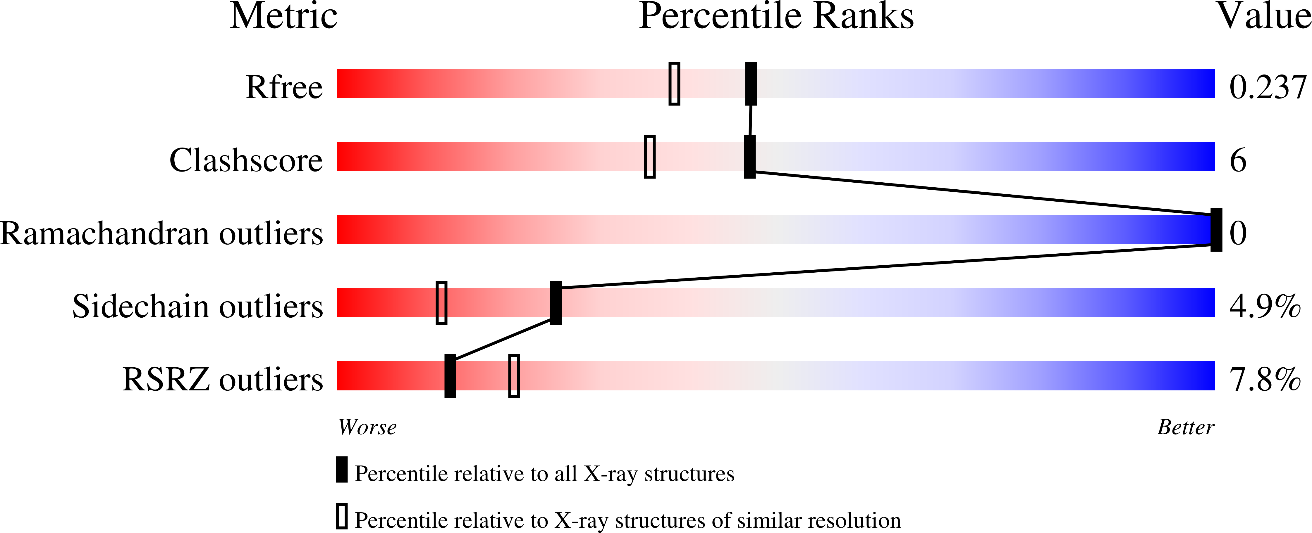
  • R-Value Free: 0.237 
  • R-Value Work: 0.194 
  • R-Value Observed: 0.197 

Starting Model: experimental
View more details

wwPDB Validation   3D Report Full Report


This is version 1.2 of the entry. See complete history


Literature

Mechanism of somatic hypermutation at the WA motif by human DNA polymerase eta.

Zhao, Y.Gregory, M.T.Biertumpfel, C.Hua, Y.J.Hanaoka, F.Yang, W.

(2013) Proc Natl Acad Sci U S A 110: 8146-8151

  • DOI: https://doi.org/10.1073/pnas.1303126110
  • Primary Citation of Related Structures:  
    4J9K, 4J9L, 4J9M, 4J9N, 4J9O, 4J9P, 4J9Q, 4J9R, 4J9S

  • PubMed Abstract: 

    Somatic hypermutation is programmed base substitutions in the variable regions of Ig genes for high-affinity antibody generation. Two motifs, RGYW and WA (R, purine; Y, pyrimidine; W, A or T), have been found to be somatic hypermutation hotspots. Overwhelming evidence suggests that DNA polymerase η (Pol η) is responsible for converting the WA motif to WG by misincorporating dGTP opposite the templating T. To elucidate the molecular mechanism, crystal structures and kinetics of human Pol η substituting dGTP for dATP in four sequence contexts, TA, AA, GA, and CA, have been determined and compared. The T:dGTP wobble base pair is stabilized by Gln-38 and Arg-61, two uniquely conserved residues among Pol η. Weak base paring of the W (T:A or A:T) at the primer end and their distinct interactions with Pol η lead to misincorporation of G in the WA motif. Between two WA motifs, our kinetic and structural data indicate that A-to-G mutation occurs more readily in the TA context than AA. Finally, Pol η can extend the T:G mispair efficiently to complete the mutagenesis.


  • Organizational Affiliation

    Institute of Nuclear-Agricultural Sciences, Zhejiang University, Hangzhou 310029, China.


Macromolecules

Find similar proteins by:  (by identity cutoff)  |  3D Structure
Entity ID: 1
MoleculeChains Sequence LengthOrganismDetailsImage
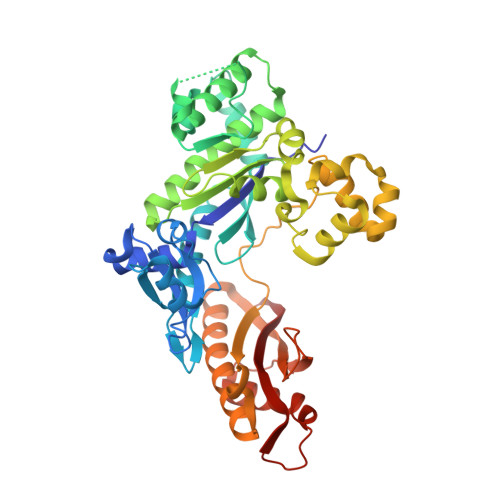
DNA polymerase eta435Homo sapiensMutation(s): 0 
Gene Names: POLHRAD30RAD30AXPV
EC: 2.7.7.7
UniProt & NIH Common Fund Data Resources
Find proteins for Q9Y253 (Homo sapiens)
Explore Q9Y253 
Go to UniProtKB:  Q9Y253
PHAROS:  Q9Y253
GTEx:  ENSG00000170734 
Entity Groups  
Sequence Clusters30% Identity50% Identity70% Identity90% Identity95% Identity100% Identity
UniProt GroupQ9Y253
Sequence Annotations
Expand
  • Reference Sequence

Find similar nucleic acids by:  Sequence   |   3D Structure  

Entity ID: 2
MoleculeChains LengthOrganismImage
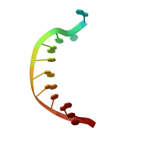
DNA (5'-D(*TP*C*AP*TP*TP*AP*TP*GP*AP*CP*GP*CP*T)-3')B [auth T]13N/A
Sequence Annotations
Expand
  • Reference Sequence

Find similar nucleic acids by:  Sequence   |   3D Structure  

Entity ID: 3
MoleculeChains LengthOrganismImage
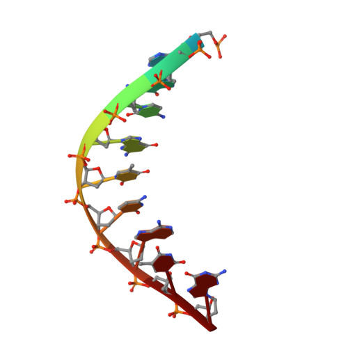
DNA (5'-D(*TP*AP*GP*CP*GP*TP*CP*AP*TP*G)-3')C [auth P]10N/A
Sequence Annotations
Expand
  • Reference Sequence
Experimental Data & Validation

Experimental Data

  • Method: X-RAY DIFFRACTION
  • Resolution: 1.96 Å
  • R-Value Free: 0.237 
  • R-Value Work: 0.194 
  • R-Value Observed: 0.197 
  • Space Group: P 61
Unit Cell:
Length ( Å )Angle ( ˚ )
a = 98.39α = 90
b = 98.39β = 90
c = 81.77γ = 120
Software Package:
Software NamePurpose
PHENIXrefinement
PDB_EXTRACTdata extraction
HKL-2000data collection
XDSdata reduction
XSCALEdata scaling
MOLREPphasing

Structure Validation

View Full Validation Report


Entry History 

Deposition Data

Revision History  (Full details and data files)

  • Version 1.0: 2013-05-01
    Type: Initial release
  • Version 1.1: 2013-08-28
    Changes: Database references
  • Version 1.2: 2023-09-20
    Changes: Data collection, Database references, Derived calculations, Refinement description