4JDC | pdb_00004jdc

Crystal structure of the Delta-pyrroline-5-carboxylate dehydrogenase from Mycobacterium tuberculosis


Experimental Data Snapshot

  • Method: X-RAY DIFFRACTION
  • Resolution: 1.60 Å
  • R-Value Free: 
    0.202 (Depositor), 0.205 (DCC) 
  • R-Value Work: 
    0.170 (Depositor), 0.179 (DCC) 
  • R-Value Observed: 
    0.172 (Depositor) 

wwPDB Validation   3D Report Full Report


Ligand Structure Quality Assessment 

Created with Raphaël 2.3.0Worse 01 BetterLigand structure goodness of fit to experimental dataBest fitted NADClick on this verticalbar to view details

This is version 1.3 of the entry. See complete history


Literature

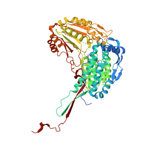
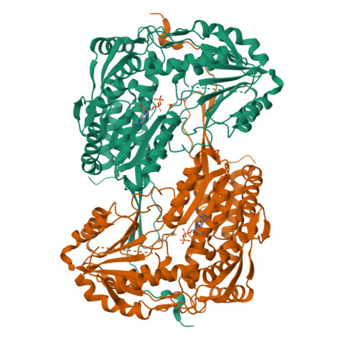
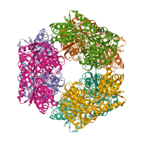
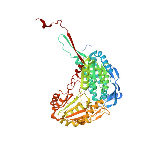
Characterization of the proline-utilization pathway in Mycobacterium tuberculosis through structural and functional studies.

Lagautriere, T.Bashiri, G.Paterson, N.G.Berney, M.Cook, G.M.Baker, E.N.

(2014) Acta Crystallogr D Biol Crystallogr 70: 968-980

  • DOI: https://doi.org/10.1107/S1399004713034391
  • Primary Citation of Related Structures:  
    4IDM, 4IDS, 4IHI, 4JDC

  • PubMed Abstract: 

    The proline-utilization pathway in Mycobacterium tuberculosis (Mtb) has recently been identified as an important factor in Mtb persistence in vivo, suggesting that this pathway could be a valuable therapeutic target against tuberculosis (TB). In Mtb, two distinct enzymes perform the conversion of proline into glutamate: the first step is the oxidation of proline into Δ(1)-pyrroline-5-carboxylic acid (P5C) by the flavoenzyme proline dehydrogenase (PruB), and the second reaction involves converting the tautomeric form of P5C (glutamate-γ-semialdehyde) into glutamate using the NAD(+)-dependent Δ(1)-pyrroline-5-carboxylic dehydrogenase (PruA). Here, the three-dimensional structures of Mtb-PruA, determined by X-ray crystallography, in the apo state and in complex with NAD(+) are described at 2.5 and 2.1 Å resolution, respectively. The structure reveals a conserved NAD(+)-binding mode, common to other related enzymes. Species-specific conformational differences in the active site, however, linked to changes in the dimer interface, suggest possibilities for selective inhibition of Mtb-PruA despite its reasonably high sequence identity to other PruA enzymes. Using recombinant PruA and PruB, the proline-utilization pathway in Mtb has also been reconstituted in vitro. Functional validation using a novel NMR approach has demonstrated that the PruA and PruB enzymes are together sufficient to convert proline to glutamate, the first such demonstration for monofunctional proline-utilization enzymes.


  • Organizational Affiliation

    Structural Biology Laboratory, School of Biological Sciences and Maurice Wilkins Centre for Molecular Biodiscovery, University of Auckland, Auckland 1010, New Zealand.


Macromolecules
Find similar proteins by:  (by identity cutoff)  |  3D Structure
Entity ID: 1
MoleculeChains Sequence LengthOrganismDetailsImage
1-pyrroline-5-carboxylate dehydrogenase563Mycobacterium tuberculosis H37RvMutation(s): 1 
Gene Names: RVBD_1187
EC: 1.5.1.12 (PDB Primary Data), 1.2.1.88 (UniProt)
UniProt
Find proteins for O50443 (Mycobacterium tuberculosis (strain ATCC 25618 / H37Rv))
Explore O50443 
Go to UniProtKB:  O50443
Entity Groups  
Sequence Clusters30% Identity50% Identity70% Identity90% Identity95% Identity100% Identity
UniProt GroupO50443
Sequence Annotations
Expand
  • Reference Sequence
Small Molecules
Ligands 1 Unique
IDChains Name / Formula / InChI Key2D Diagram3D Interactions
NAD
Query on NAD

Download Ideal Coordinates CCD File 
B [auth A]NICOTINAMIDE-ADENINE-DINUCLEOTIDE
C21 H27 N7 O14 P2
BAWFJGJZGIEFAR-NNYOXOHSSA-N
Modified Residues  1 Unique
IDChains TypeFormula2D DiagramParent
CME
Query on CME
A
L-PEPTIDE LINKINGC5 H11 N O3 S2CYS
Experimental Data & Validation

Experimental Data

  • Method: X-RAY DIFFRACTION
  • Resolution: 1.60 Å
  • R-Value Free:  0.202 (Depositor), 0.205 (DCC) 
  • R-Value Work:  0.170 (Depositor), 0.179 (DCC) 
  • R-Value Observed: 0.172 (Depositor) 
Space Group: P 6 2 2
Unit Cell:
Length ( Å )Angle ( ˚ )
a = 163.133α = 90
b = 163.133β = 90
c = 96.181γ = 120
Software Package:
Software NamePurpose
Blu-Icedata collection
MOLREPphasing
REFMACrefinement
XDSdata reduction
SCALAdata scaling

Structure Validation

View Full Validation Report


Ligand Structure Quality Assessment 

Created with Raphaël 2.3.0Worse 01 BetterLigand structure goodness of fit to experimental dataBest fitted NADClick on this verticalbar to view details

Entry History 

Deposition Data

Revision History  (Full details and data files)

  • Version 1.0: 2013-12-18
    Type: Initial release
  • Version 1.1: 2014-04-16
    Changes: Database references
  • Version 1.2: 2017-11-15
    Changes: Refinement description
  • Version 1.3: 2024-11-27
    Changes: Data collection, Database references, Derived calculations, Structure summary