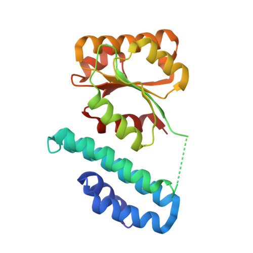
4JGI

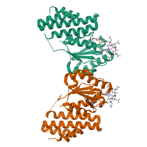
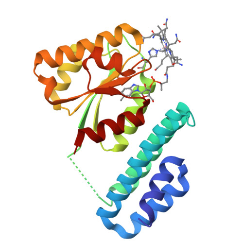
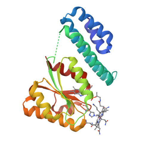
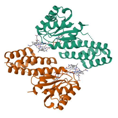
1.5 Angstrom crystal structure of a novel cobalamin-binding protein from Desulfitobacterium hafniense DCB-2


Experimental Data Snapshot

  • Method: X-RAY DIFFRACTION
  • Resolution: 1.50 Å
  • R-Value Free: 0.194 
  • R-Value Work: 0.147 
  • R-Value Observed: 0.150 

wwPDB Validation   3D Report Full Report


Ligand Structure Quality Assessment 


This is version 1.2 of the entry. See complete history


Literature

Structure of the cobalamin-binding protein of a putative O-demethylase from Desulfitobacterium hafniense DCB-2.

Sjuts, H.Dunstan, M.S.Fisher, K.Leys, D.

(2013) Acta Crystallogr D Biol Crystallogr 69: 1609-1616

  • DOI: https://doi.org/10.1107/S0907444913011323
  • Primary Citation of Related Structures:  
    4JGI

  • PubMed Abstract: 

    This study describes the identification and the structural and spectroscopic analysis of a cobalamin-binding protein (termed CobDH) implicated in O-demethylation by the organohalide-respiring bacterium Desulfitobacterium hafniense DCB-2. The 1.5 Å resolution crystal structure of CobDH is presented in the cobalamin-bound state and reveals that the protein is composed of an N-terminal helix-bundle domain and a C-terminal Rossmann-fold domain, with the cobalamin coordinated in the base-off/His-on conformation similar to other cobalamin-binding domains that catalyse methyl-transfer reactions. EPR spectroscopy of CobDH confirms cobalamin binding and reveals the presence of a cob(III)alamin superoxide, indicating binding of oxygen to the fully oxidized cofactor. These data provide the first structural insights into the methyltransferase reactions that occur during O-demethylation by D. hafniense.


  • Organizational Affiliation

    Manchester Institute of Biotechnology, Faculty of Life Sciences, University of Manchester, 131 Princess Street, Manchester M1 7DN, England.


Macromolecules
Find similar proteins by:  (by identity cutoff)  |  3D Structure
Entity ID: 1
MoleculeChains Sequence LengthOrganismDetailsImage
Putative uncharacterized protein
A, B
211Desulfitobacterium hafnienseMutation(s): 0 
UniProt
Find proteins for Q8RPG0 (Desulfitobacterium hafniense)
Explore Q8RPG0 
Go to UniProtKB:  Q8RPG0
Entity Groups  
Sequence Clusters30% Identity50% Identity70% Identity90% Identity95% Identity100% Identity
UniProt GroupQ8RPG0
Sequence Annotations
Expand
  • Reference Sequence
Small Molecules
Ligands 1 Unique
IDChains Name / Formula / InChI Key2D Diagram3D Interactions
COB
Query on COB

Download Ideal Coordinates CCD File 
C [auth A],
D [auth B]
CO-METHYLCOBALAMIN
C63 H91 Co N13 O14 P
BMOGZGJBQIUQJG-BYLSPCLQSA-N
Experimental Data & Validation

Experimental Data

  • Method: X-RAY DIFFRACTION
  • Resolution: 1.50 Å
  • R-Value Free: 0.194 
  • R-Value Work: 0.147 
  • R-Value Observed: 0.150 
  • Space Group: P 31
Unit Cell:
Length ( Å )Angle ( ˚ )
a = 69.99α = 90
b = 69.99β = 90
c = 91.68γ = 120
Software Package:
Software NamePurpose
ADSCdata collection
PHASERphasing
REFMACrefinement
xia2data reduction
xia2data scaling

Structure Validation

View Full Validation Report


Ligand Structure Quality Assessment 


Entry History 

Deposition Data

Revision History  (Full details and data files)

  • Version 1.0: 2013-08-07
    Type: Initial release
  • Version 1.1: 2017-11-15
    Changes: Refinement description
  • Version 1.2: 2024-02-28
    Changes: Data collection, Database references, Derived calculations