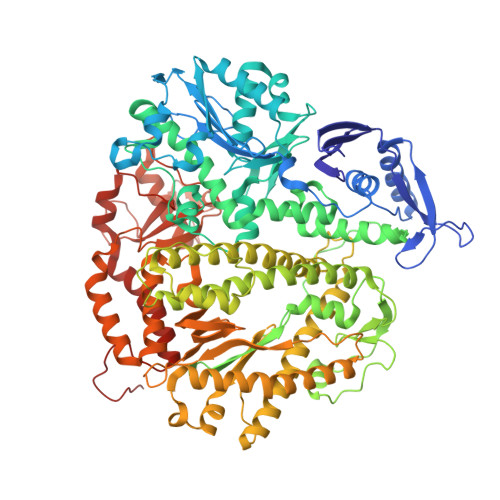
4KHN

Crystal structure of the ternary complex of the D714A mutant of RB69 DNA polymerase


Experimental Data Snapshot

  • Method: X-RAY DIFFRACTION
  • Resolution: 2.55 Å
  • R-Value Free: 0.211 
  • R-Value Work: 0.168 
  • R-Value Observed: 0.170 

Starting Model: experimental
View more details

wwPDB Validation   3D Report Full Report


Ligand Structure Quality Assessment 


This is version 1.2 of the entry. See complete history


Literature

A Remote Palm Domain Residue of RB69 DNA Polymerase Is Critical for Enzyme Activity and Influences the Conformation of the Active Site.

Jacewicz, A.Trzemecka, A.Guja, K.E.Plochocka, D.Yakubovskaya, E.Bebenek, A.Garcia-Diaz, M.

(2013) PLoS One 8: e76700-e76700

  • DOI: https://doi.org/10.1371/journal.pone.0076700
  • Primary Citation of Related Structures:  
    4I9L, 4I9Q, 4KHN

  • PubMed Abstract: 

    Non-conserved amino acids that are far removed from the active site can sometimes have an unexpected effect on enzyme catalysis. We have investigated the effects of alanine replacement of residues distant from the active site of the replicative RB69 DNA polymerase, and identified a substitution in a weakly conserved palm residue (D714A), that renders the enzyme incapable of sustaining phage replication in vivo. D714, located several angstroms away from the active site, does not contact the DNA or the incoming dNTP, and our apoenzyme and ternary crystal structures of the Pol(D714A) mutant demonstrate that D714A does not affect the overall structure of the protein. The structures reveal a conformational change of several amino acid side chains, which cascade out from the site of the substitution towards the catalytic center, substantially perturbing the geometry of the active site. Consistent with these structural observations, the mutant has a significantly reduced k pol for correct incorporation. We propose that the observed structural changes underlie the severe polymerization defect and thus D714 is a remote, non-catalytic residue that is nevertheless critical for maintaining an optimal active site conformation. This represents a striking example of an action-at-a-distance interaction.


  • Organizational Affiliation

    Department of Molecular Biology, Institute of Biochemistry and Biophysics, Polish Academy of Sciences, Warsaw, Poland.


Macromolecules

Find similar proteins by:  (by identity cutoff)  |  3D Structure
Entity ID: 1
MoleculeChains Sequence LengthOrganismDetailsImage
DNA polymeraseA,
F [auth B]
903Escherichia phage RB69Mutation(s): 4 
Gene Names: 43
EC: 2.7.7.7 (PDB Primary Data), 3.1.11 (UniProt)
UniProt
Find proteins for Q38087 (Escherichia phage RB69)
Explore Q38087 
Go to UniProtKB:  Q38087
Entity Groups  
Sequence Clusters30% Identity50% Identity70% Identity90% Identity95% Identity100% Identity
UniProt GroupQ38087
Sequence Annotations
Expand
  • Reference Sequence

Find similar nucleic acids by:  Sequence   |   3D Structure  

Entity ID: 2
MoleculeChains LengthOrganismImage
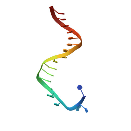
DNA (5'-D(*TP*CP*AP*CP*GP*TP*AP*AP*GP*CP*AP*GP*TP*CP*CP*GP*CP*G)-3')B [auth C],
D [auth E]
18N/A
Sequence Annotations
Expand
  • Reference Sequence

Find similar nucleic acids by:  Sequence   |   3D Structure  

Entity ID: 3
MoleculeChains LengthOrganismImage
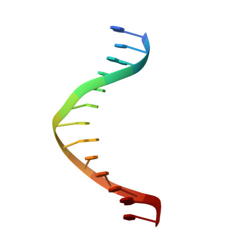
DNA (5'-D(*GP*CP*GP*GP*AP*CP*TP*GP*CP*TP*TP*AP*C)-3')C [auth D],
E [auth F]
13N/A
Sequence Annotations
Expand
  • Reference Sequence
Small Molecules
Ligands 5 Unique
IDChains Name / Formula / InChI Key2D Diagram3D Interactions
XG4
Query on XG4

Download Ideal Coordinates CCD File 
G [auth A],
Q [auth C],
R [auth B],
S [auth B]
2'-deoxy-5'-O-[(R)-hydroxy{[(R)-hydroxy(phosphonooxy)phosphoryl]amino}phosphoryl]guanosine
C10 H17 N6 O12 P3
DWGAAFQEGIMTIA-KVQBGUIXSA-N
SO4
Query on SO4

Download Ideal Coordinates CCD File 
I [auth A]
J [auth A]
K [auth A]
L [auth A]
M [auth A]
I [auth A],
J [auth A],
K [auth A],
L [auth A],
M [auth A],
T [auth B]
SULFATE ION
O4 S
QAOWNCQODCNURD-UHFFFAOYSA-L
GOL
Query on GOL

Download Ideal Coordinates CCD File 
N [auth A],
O [auth A]
GLYCEROL
C3 H8 O3
PEDCQBHIVMGVHV-UHFFFAOYSA-N
CA
Query on CA

Download Ideal Coordinates CCD File 
H [auth A],
U [auth B]
CALCIUM ION
Ca
BHPQYMZQTOCNFJ-UHFFFAOYSA-N
NA
Query on NA

Download Ideal Coordinates CCD File 
P [auth A]SODIUM ION
Na
FKNQFGJONOIPTF-UHFFFAOYSA-N
Experimental Data & Validation

Experimental Data

  • Method: X-RAY DIFFRACTION
  • Resolution: 2.55 Å
  • R-Value Free: 0.211 
  • R-Value Work: 0.168 
  • R-Value Observed: 0.170 
  • Space Group: P 1 21 1
Unit Cell:
Length ( Å )Angle ( ˚ )
a = 74.258α = 90
b = 119.353β = 91.64
c = 148.031γ = 90
Software Package:
Software NamePurpose
CBASSdata collection
PHASERphasing
PHENIXrefinement
XDSdata reduction
SCALAdata scaling

Structure Validation

View Full Validation Report


Ligand Structure Quality Assessment 


Entry History 

Deposition Data

Revision History  (Full details and data files)

  • Version 1.0: 2013-10-09
    Type: Initial release
  • Version 1.1: 2013-10-23
    Changes: Database references
  • Version 1.2: 2023-09-20
    Changes: Data collection, Database references, Derived calculations, Refinement description