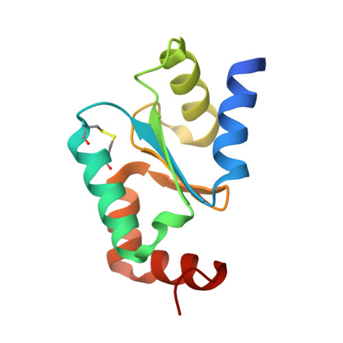
4MZB

Crystal structure of Grx1 from Plasmodium falciparum


Experimental Data Snapshot

  • Method: X-RAY DIFFRACTION
  • Resolution: 1.04 Å
  • R-Value Free: 0.129 
  • R-Value Work: 0.117 
  • R-Value Observed: 0.118 

Starting Model: experimental
View more details

wwPDB Validation   3D Report Full Report


Ligand Structure Quality Assessment 


This is version 1.4 of the entry. See complete history


Literature

Atomic resolution crystal structure of glutaredoxin 1 from Plasmodium falciparum and comparison with other glutaredoxins.

Yogavel, M.Tripathi, T.Gupta, A.Banday, M.M.Rahlfs, S.Becker, K.Belrhali, H.Sharma, A.

(2014) Acta Crystallogr D Biol Crystallogr 70: 91-100

  • DOI: https://doi.org/10.1107/S1399004713025285
  • Primary Citation of Related Structures:  
    4HJM, 4MZB, 4MZC

  • PubMed Abstract: 

    Glutaredoxins (Grxs) are redox proteins that use glutathione ((γ)Glu-Cys-Gly; GSH) as a cofactor. Plasmodium falciparum has one classic dithiol (CXXC) glutaredoxin (glutaredoxin 1; PfGrx1) and three monothiol (CXXS) Grx-like proteins (GLPs), which have five residue insertions prior to the active-site Cys. Here, the crystal structure of PfGrx1 has been determined by the sulfur single-wavelength anomalous diffraction (S-SAD) method utilizing intrinsic protein and solvent S atoms. Several residues were modelled with alternate conformations, and an alternate position was refined for the active-site Cys29 owing to radiation damage. The GSH-binding site is occupied by water polygons and buffer molecules. Structural comparison of PfGrx1 with other Grxs and Grx-like proteins revealed that the GSH-binding motifs (CXXC/CXXS, TVP, CDD, Lys26 and Gln/Arg63) are structurally conserved. Both the monothiol and dithiol Grxs possess three conserved water molecules; two of these were located in the GSH-binding site. PfGrx1 has several polar and charged amino-acid substitutions that provide structurally important additional hydrogen bonds and salt bridges missing in other Grxs.


  • Organizational Affiliation

    Structural and Computational Biology Group, International Centre for Genetic Engineering and Biotechnology (ICGEB), Aruna Asaf Ali Road, New Delhi 110 067, India.


Macromolecules
Find similar proteins by:  (by identity cutoff)  |  3D Structure
Entity ID: 1
MoleculeChains Sequence LengthOrganismDetailsImage
Glutaredoxin111Plasmodium falciparum 3D7Mutation(s): 0 
Gene Names: GRX1PFC0271c
EC: 1.20.4.1
UniProt
Find proteins for Q9NLB2 (Plasmodium falciparum (isolate 3D7))
Explore Q9NLB2 
Go to UniProtKB:  Q9NLB2
Entity Groups  
Sequence Clusters30% Identity50% Identity70% Identity90% Identity95% Identity100% Identity
UniProt GroupQ9NLB2
Sequence Annotations
Expand
  • Reference Sequence
Small Molecules
Experimental Data & Validation

Experimental Data

  • Method: X-RAY DIFFRACTION
  • Resolution: 1.04 Å
  • R-Value Free: 0.129 
  • R-Value Work: 0.117 
  • R-Value Observed: 0.118 
  • Space Group: P 32 2 1
Unit Cell:
Length ( Å )Angle ( ˚ )
a = 47.953α = 90
b = 47.953β = 90
c = 82.45γ = 120
Software Package:
Software NamePurpose
MOLREPphasing
PHENIXrefinement
PDB_EXTRACTdata extraction
DNAdata collection
HKL-2000data reduction
HKL-2000data scaling

Structure Validation

View Full Validation Report


Ligand Structure Quality Assessment 


Entry History 

Deposition Data

Revision History  (Full details and data files)

  • Version 1.0: 2013-10-09
    Type: Initial release
  • Version 1.1: 2014-01-08
    Changes: Database references
  • Version 1.2: 2014-09-24
    Changes: Database references
  • Version 1.3: 2020-09-09
    Changes: Derived calculations, Structure summary
  • Version 1.4: 2023-09-20
    Changes: Data collection, Database references, Refinement description