4NC7 | pdb_00004nc7

N-terminal domain of delta-subunit of RNA polymerase complexed with I3C and nickel ions


Experimental Data Snapshot

  • Method: X-RAY DIFFRACTION
  • Resolution: 2.00 Å
  • R-Value Free: 
    0.259 (Depositor), 0.260 (DCC) 
  • R-Value Work: 
    0.201 (Depositor), 0.210 (DCC) 
  • R-Value Observed: 
    0.204 (Depositor) 

wwPDB Validation   3D Report Full Report


Ligand Structure Quality Assessment 

Created with Raphaël 2.3.0Worse 01 BetterLigand structure goodness of fit to experimental dataBest fitted I3CClick on this verticalbar to view details

This is version 1.2 of the entry. See complete history


Literature

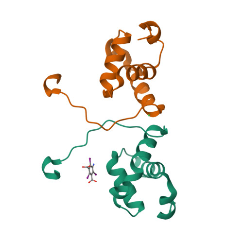
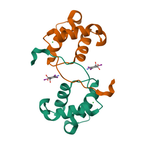
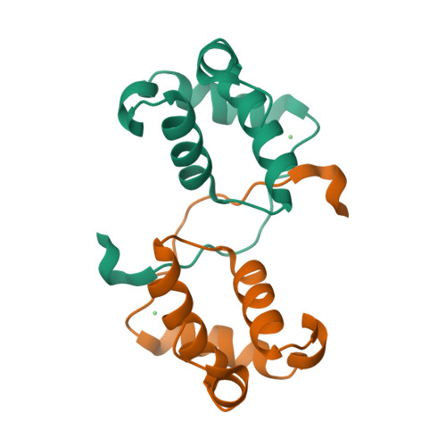
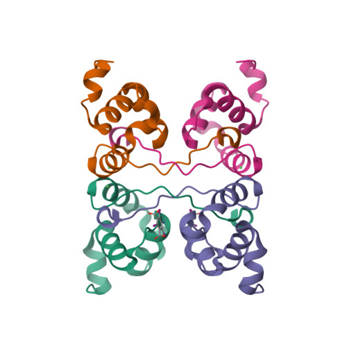
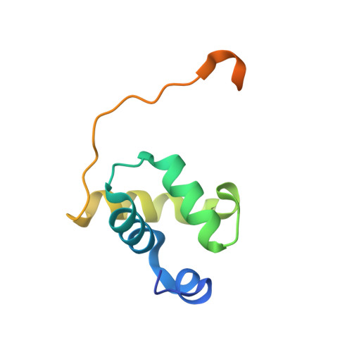
X-ray vs. NMR structure of N-terminal domain of delta-subunit of RNA polymerase.

Demo, G.Papouskova, V.Komarek, J.Kaderavek, P.Otrusinova, O.Srb, P.Rabatinova, A.Krasny, L.Zidek, L.Sklenar, V.Wimmerova, M.

(2014) J Struct Biol 187: 174-186

  • DOI: https://doi.org/10.1016/j.jsb.2014.06.001
  • Primary Citation of Related Structures:  
    4NC7, 4NC8

  • PubMed Abstract: 

    The crystal structure of the N-terminal domain of the RNA polymerase δ subunit (Nδ) from Bacillus subtilis solved at a resolution of 2.0Å is compared with the NMR structure determined previously. The molecule crystallizes in the space group C222(1) with a dimer in the asymmetric unit. Importantly, the X-ray structure exhibits significant differences from the lowest energy NMR structure. In addition to the overall structure differences, structurally important β sheets found in the NMR structure are not present in the crystal structure. We systematically investigated the cause of the discrepancies between the NMR and X-ray structures of Nδ, addressing the pH dependence, presence of metal ions, and crystal packing forces. We convincingly showed that the crystal packing forces, together with the presence of Ni(2+) ions, are the main reason for such a difference. In summary, the study illustrates that the two structural approaches may give unequal results, which need to be interpreted with care to obtain reliable structural information in terms of biological relevance.


  • Organizational Affiliation

    National Centre for Biomolecular Research, Faculty of Science, Masaryk University, Kamenice 5/A4, 62500 Brno, Czech Republic; Central European Institute of Technology-CEITEC, Masaryk University, Kamenice 5/A4, 62500 Brno, Czech Republic.


Macromolecules
Find similar proteins by:  (by identity cutoff)  |  3D Structure
Entity ID: 1
MoleculeChains Sequence LengthOrganismDetailsImage
DNA-directed RNA polymerase subunit delta
A, B
99Bacillus subtilis subsp. subtilis str. 168Mutation(s): 0 
Gene Names: rpoEBSU37160
UniProt
Find proteins for P12464 (Bacillus subtilis (strain 168))
Explore P12464 
Go to UniProtKB:  P12464
Entity Groups  
Sequence Clusters30% Identity50% Identity70% Identity90% Identity95% Identity100% Identity
UniProt GroupP12464
Sequence Annotations
Expand
  • Reference Sequence
Experimental Data & Validation

Experimental Data

  • Method: X-RAY DIFFRACTION
  • Resolution: 2.00 Å
  • R-Value Free:  0.259 (Depositor), 0.260 (DCC) 
  • R-Value Work:  0.201 (Depositor), 0.210 (DCC) 
  • R-Value Observed: 0.204 (Depositor) 
Space Group: C 2 2 21
Unit Cell:
Length ( Å )Angle ( ˚ )
a = 41.65α = 90
b = 110.2β = 90
c = 85.26γ = 90
Software Package:
Software NamePurpose
MAR345dtbdata collection
Auto-Rickshawphasing
MOLREPphasing
REFMACrefinement
MOSFLMdata reduction
SCALAdata scaling

Structure Validation

View Full Validation Report


Ligand Structure Quality Assessment 

Created with Raphaël 2.3.0Worse 01 BetterLigand structure goodness of fit to experimental dataBest fitted I3CClick on this verticalbar to view details

Entry History 

Deposition Data

Revision History  (Full details and data files)

  • Version 1.0: 2014-07-02
    Type: Initial release
  • Version 1.1: 2014-08-20
    Changes: Database references
  • Version 1.2: 2024-02-28
    Changes: Data collection, Database references, Derived calculations