4Q01

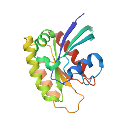
Second-site screening of K-Ras in the presence of covalently attached first-site ligands


Experimental Data Snapshot

  • Method: X-RAY DIFFRACTION
  • Resolution: 1.29 Å
  • R-Value Free: 0.213 
  • R-Value Work: 0.188 
  • R-Value Observed: 0.189 

wwPDB Validation   3D Report Full Report


Ligand Structure Quality Assessment 


This is version 1.1 of the entry. See complete history


Literature

A method for the second-site screening of K-Ras in the presence of a covalently attached first-site ligand.

Sun, Q.Phan, J.Friberg, A.R.Camper, D.V.Olejniczak, E.T.Fesik, S.W.

(2014) J Biomol NMR 60: 11-14

  • DOI: https://doi.org/10.1007/s10858-014-9849-8
  • Primary Citation of Related Structures:  
    4PZY, 4PZZ, 4Q01, 4Q02, 4Q03

  • PubMed Abstract: 

    K-Ras is a well-validated cancer target but is considered to be "undruggable" due to the lack of suitable binding pockets. We previously discovered small molecules that bind weakly to K-Ras but wanted to improve their binding affinities by identifying ligands that bind near our initial hits that we could link together. Here we describe an approach for identifying second site ligands that uses a cysteine residue to covalently attach a compound for tight binding to the first site pocket followed by a fragment screen for binding to a second site. This approach could be very useful for targeting Ras and other challenging drug targets.


  • Organizational Affiliation

    Department of Biochemistry, Vanderbilt University School of Medicine, Nashville, TN, 37232-0146, USA.


Macromolecules
Find similar proteins by:  (by identity cutoff)  |  3D Structure
Entity ID: 1
MoleculeChains Sequence LengthOrganismDetailsImage
K-Ras
A, B
169Homo sapiensMutation(s): 3 
EC: 3.6.5.2
UniProt & NIH Common Fund Data Resources
Find proteins for P01116 (Homo sapiens)
Explore P01116 
Go to UniProtKB:  P01116
PHAROS:  P01116
GTEx:  ENSG00000133703 
Entity Groups  
Sequence Clusters30% Identity50% Identity70% Identity90% Identity95% Identity100% Identity
UniProt GroupP01116
Sequence Annotations
Expand
  • Reference Sequence
Experimental Data & Validation

Experimental Data

  • Method: X-RAY DIFFRACTION
  • Resolution: 1.29 Å
  • R-Value Free: 0.213 
  • R-Value Work: 0.188 
  • R-Value Observed: 0.189 
  • Space Group: P 21 21 21
Unit Cell:
Length ( Å )Angle ( ˚ )
a = 36.314α = 90
b = 83.671β = 90
c = 104.835γ = 90
Software Package:
Software NamePurpose
PROTEUM PLUSdata collection
PHENIXmodel building
PHENIXrefinement
HKL-2000data reduction
HKL-2000data scaling
PHENIXphasing

Structure Validation

View Full Validation Report


Ligand Structure Quality Assessment 


Entry History 

Deposition Data

Revision History  (Full details and data files)

  • Version 1.0: 2014-09-10
    Type: Initial release
  • Version 1.1: 2024-11-27
    Changes: Data collection, Database references, Derived calculations, Structure summary