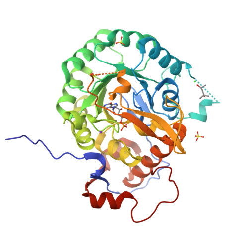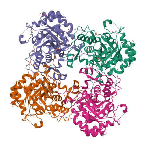
Crystal Structure of Inosine 5'-monophosphate Dehydrogenase with the Internal Deletion Containing CBS Domain from Campylobacter jejuni
Kim, Y., Makowska-Grzyska, M., Gu, M., Hedstrom, L., Anderson, W.F., Joachimiak, A., CSGIDTo be published.
Experimental Data Snapshot
Starting Model: experimental
View more details
Entity ID: 1 | |||||
|---|---|---|---|---|---|
| Molecule | Chains | Sequence Length | Organism | Details | Image |
| Inosine-5'-monophosphate dehydrogenase | 385 | Campylobacter jejuni subsp. jejuni NCTC 11168 = ATCC 700819 | Mutation(s): 0 Gene Names: guaB, Cj1058c EC: 1.1.1.205 | 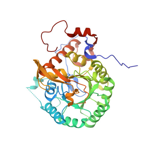 | |
UniProt | |||||
Find proteins for Q0P9J4 (Campylobacter jejuni subsp. jejuni serotype O:2 (strain ATCC 700819 / NCTC 11168)) Explore Q0P9J4 Go to UniProtKB: Q0P9J4 | |||||
Entity Groups | |||||
| Sequence Clusters | 30% Identity50% Identity70% Identity90% Identity95% Identity100% Identity | ||||
| UniProt Group | Q0P9J4 | ||||
Sequence AnnotationsExpand | |||||
| |||||
| Ligands 6 Unique | |||||
|---|---|---|---|---|---|
| ID | Chains | Name / Formula / InChI Key | 2D Diagram | 3D Interactions | |
| IMP Query on IMP | B [auth A] | INOSINIC ACID C10 H13 N4 O8 P GRSZFWQUAKGDAV-KQYNXXCUSA-N |  | ||
| MPD Query on MPD | H [auth A] | (4S)-2-METHYL-2,4-PENTANEDIOL C6 H14 O2 SVTBMSDMJJWYQN-YFKPBYRVSA-N |  | ||
| SO4 Query on SO4 | E [auth A], F [auth A], G [auth A] | SULFATE ION O4 S QAOWNCQODCNURD-UHFFFAOYSA-L |  | ||
| GOL Query on GOL | D [auth A] | GLYCEROL C3 H8 O3 PEDCQBHIVMGVHV-UHFFFAOYSA-N |  | ||
| K Query on K | I [auth A] | POTASSIUM ION K NPYPAHLBTDXSSS-UHFFFAOYSA-N |  | ||
| CL Query on CL | C [auth A] | CHLORIDE ION Cl VEXZGXHMUGYJMC-UHFFFAOYSA-M |  | ||
| Length ( Å ) | Angle ( ˚ ) |
|---|---|
| a = 119.413 | α = 90 |
| b = 119.413 | β = 90 |
| c = 69.394 | γ = 90 |
| Software Name | Purpose |
|---|---|
| SBC-Collect | data collection |
| HKL-3000 | data collection |
| HKL-3000 | phasing |
| MOLREP | phasing |
| PHENIX | refinement |
| HKL-3000 | data reduction |
| HKL-3000 | data scaling |
RCSB PDB is hosted by

RCSB PDB is a member of the