4RJT

Crystal Structure of Unliganded, Full Length hUGDH at pH 7.0


Experimental Data Snapshot

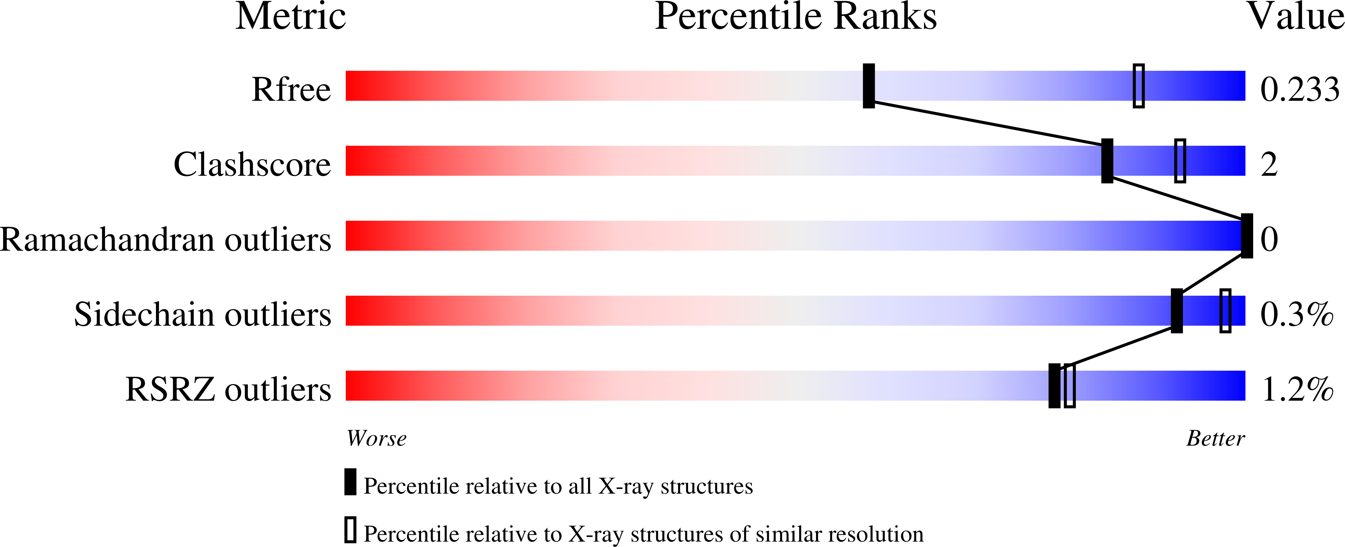
  • Method: X-RAY DIFFRACTION
  • Resolution: 2.70 Å
  • R-Value Free: 0.231 
  • R-Value Work: 0.183 
  • R-Value Observed: 0.185 

Starting Model: experimental
View more details

wwPDB Validation   3D Report Full Report


This is version 1.3 of the entry. See complete history


Literature

Hysteresis in Human UDP-Glucose Dehydrogenase Is Due to a Restrained Hexameric Structure That Favors Feedback Inhibition.

Kadirvelraj, R.Custer, G.S.Keul, N.D.Sennett, N.C.Sidlo, A.M.Walsh Jr., R.M.Wood, Z.A.

(2014) Biochemistry 53: 8043-8051

  • DOI: https://doi.org/10.1021/bi500594x
  • Primary Citation of Related Structures:  
    4RJT

  • PubMed Abstract: 

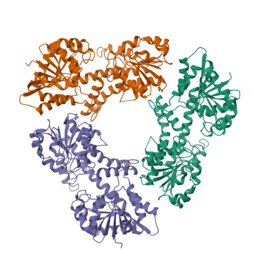
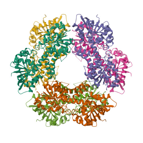
    Human UDP-α-d-glucose-6-dehydrogenase (hUGDH) displays hysteresis because of a slow isomerization from an inactive state (E*) to an active state (E). Here we show that the structure of E* constrains hUGDH in a conformation that favors feedback inhibition at physiological pH. The feedback inhibitor UDP-α-d-xylose (UDP-Xyl) competes with the substrate UDP-α-d-glucose for the active site. Upon binding, UDP-Xyl triggers an allosteric switch that changes the structure and affinity of the intersubunit interface to form a stable but inactive horseshoe-shaped hexamer. Using sedimentation velocity studies and a new crystal structure, we show that E* represents a stable conformational intermediate between the active and feedback-inhibited conformations. Because the allosteric switch occludes the cofactor and substrate binding sites in the inactive hexamer, the intermediate conformation observed in the crystal structure is consistent with the E* transient observed in relaxation studies. Steady-state analysis shows that the E* conformation enhances the affinity of hUGDH for the allosteric inhibitor UDP-Xyl by 8.6-fold (Ki = 810 nM). We present a model in which the constrained quaternary structure permits a small effector molecule to leverage a disproportionately large allosteric response.


  • Organizational Affiliation

    Department of Biochemistry & Molecular Biology, University of Georgia , Athens, Georgia 30602, United States.


Macromolecules
Find similar proteins by:  (by identity cutoff)  |  3D Structure
Entity ID: 1
MoleculeChains Sequence LengthOrganismDetailsImage
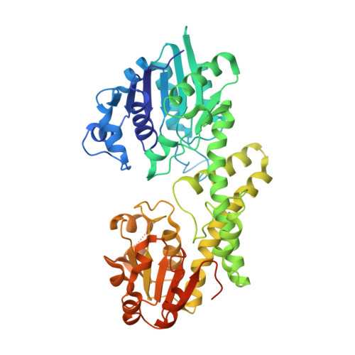
UDP-glucose 6-dehydrogenase
A, B, C
494Homo sapiensMutation(s): 0 
Gene Names: UGDH
EC: 1.1.1.22
UniProt & NIH Common Fund Data Resources
Find proteins for O60701 (Homo sapiens)
Explore O60701 
Go to UniProtKB:  O60701
PHAROS:  O60701
GTEx:  ENSG00000109814 
Entity Groups  
Sequence Clusters30% Identity50% Identity70% Identity90% Identity95% Identity100% Identity
UniProt GroupO60701
Sequence Annotations
Expand
  • Reference Sequence
Experimental Data & Validation

Experimental Data

  • Method: X-RAY DIFFRACTION
  • Resolution: 2.70 Å
  • R-Value Free: 0.231 
  • R-Value Work: 0.183 
  • R-Value Observed: 0.185 
  • Space Group: C 1 2 1
Unit Cell:
Length ( Å )Angle ( ˚ )
a = 176.98α = 90
b = 113.96β = 116.91
c = 97.12γ = 90
Software Package:
Software NamePurpose
XDSdata scaling
PHENIXmodel building
PHENIXrefinement
XDSdata reduction
XSCALEdata scaling
PHENIXphasing

Structure Validation

View Full Validation Report


Entry History 

Deposition Data

Revision History  (Full details and data files)

  • Version 1.0: 2014-12-31
    Type: Initial release
  • Version 1.1: 2015-01-21
    Changes: Database references
  • Version 1.2: 2018-04-25
    Changes: Data collection, Database references
  • Version 1.3: 2023-09-20
    Changes: Data collection, Database references, Derived calculations, Refinement description