4U9N | pdb_00004u9n

Structure of a membrane protein


Experimental Data Snapshot

  • Method: X-RAY DIFFRACTION
  • Resolution: 2.20 Å
  • R-Value Free: 
    0.240 (Depositor), 0.240 (DCC) 
  • R-Value Work: 
    0.224 (Depositor), 0.230 (DCC) 
  • R-Value Observed: 
    0.224 (Depositor) 

wwPDB Validation   3D Report Full Report


Ligand Structure Quality Assessment 

Created with Raphaël 2.3.0Worse 01 BetterLigand structure goodness of fit to experimental dataBest fitted OLCClick on this verticalbar to view detailsBest fitted OLAClick on this verticalbar to view details

This is version 1.2 of the entry. See complete history


Literature

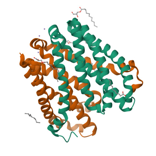
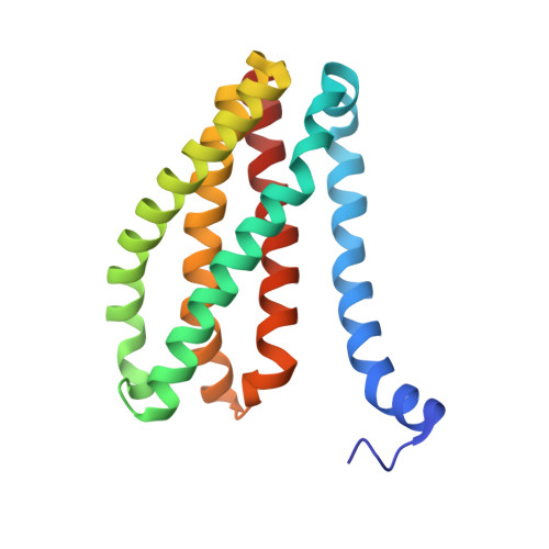
Structural basis for ion selectivity revealed by high-resolution crystal structure of Mg(2+) channel MgtE

Takeda, H.Hattori, M.Nishizawa, T.Yamashita, K.Shah, S.T.Caffrey, M.Maturana, A.D.Ishitani, R.Nureki, O.

(2014) Nat Commun 5: 5374-5374

  • DOI: https://doi.org/10.1038/ncomms6374
  • Primary Citation of Related Structures:  
    4U9L, 4U9N, 4WIB

  • PubMed Abstract: 

    Magnesium is the most abundant divalent cation in living cells and is crucial to several biological processes. MgtE is a Mg(2+) channel distributed in all domains of life that contributes to the maintenance of cellular Mg(2+) homeostasis. Here we report the high-resolution crystal structures of the transmembrane domain of MgtE, bound to Mg(2+), Mn(2+) and Ca(2+). The high-resolution Mg(2+)-bound crystal structure clearly visualized the hydrated Mg(2+) ion within its selectivity filter. Based on those structures and biochemical analyses, we propose a cation selectivity mechanism for MgtE in which the geometry of the hydration shell of the fully hydrated Mg(2+) ion is recognized by the side-chain carboxylate groups in the selectivity filter. This is in contrast to the K(+)-selective filter of KcsA, which recognizes a dehydrated K(+) ion. Our results further revealed a cation-binding site on the periplasmic side, which regulate channel opening and prevents conduction of near-cognate cations.


  • Organizational Affiliation

    1] Department of Biological Sciences, Graduate School of Science, University of Tokyo, 2-11-16 Yayoi, Bunkyo-ku, Tokyo 113-0032, Japan [2] Global Research Cluster, RIKEN, 2-1 Hirosawa, Wako-shi, Saitama 351-0198, Japan.


Macromolecules
Find similar proteins by:  (by identity cutoff)  |  3D Structure
Entity ID: 1
MoleculeChains Sequence LengthOrganismDetailsImage
Magnesium transporter MgtE
A, B
178Thermus thermophilus HB8Mutation(s): 0 
Gene Names: TTHA1060
Membrane Entity: Yes 
UniProt
Find proteins for Q5SMG8 (Thermus thermophilus (strain ATCC 27634 / DSM 579 / HB8))
Explore Q5SMG8 
Go to UniProtKB:  Q5SMG8
Entity Groups  
Sequence Clusters30% Identity50% Identity70% Identity90% Identity95% Identity100% Identity
UniProt GroupQ5SMG8
Sequence Annotations
Expand
  • Reference Sequence
Small Molecules
Ligands 3 Unique
IDChains Name / Formula / InChI Key2D Diagram3D Interactions
OLC
Query on OLC

Download Ideal Coordinates CCD File 
F [auth A],
G [auth A],
K [auth B],
L [auth B],
M [auth B]
(2R)-2,3-dihydroxypropyl (9Z)-octadec-9-enoate
C21 H40 O4
RZRNAYUHWVFMIP-GDCKJWNLSA-N
OLA
Query on OLA

Download Ideal Coordinates CCD File 
E [auth A],
J [auth B]
OLEIC ACID
C18 H34 O2
ZQPPMHVWECSIRJ-KTKRTIGZSA-N
MN
Query on MN

Download Ideal Coordinates CCD File 
C [auth A],
D [auth A],
H [auth B],
I [auth B]
MANGANESE (II) ION
Mn
WAEMQWOKJMHJLA-UHFFFAOYSA-N
Experimental Data & Validation

Experimental Data

  • Method: X-RAY DIFFRACTION
  • Resolution: 2.20 Å
  • R-Value Free:  0.240 (Depositor), 0.240 (DCC) 
  • R-Value Work:  0.224 (Depositor), 0.230 (DCC) 
  • R-Value Observed: 0.224 (Depositor) 
Space Group: P 21 21 21
Unit Cell:
Length ( Å )Angle ( ˚ )
a = 63.521α = 90
b = 70.681β = 90
c = 103.327γ = 90
Software Package:
Software NamePurpose
PHENIXrefinement

Structure Validation

View Full Validation Report


Ligand Structure Quality Assessment 

Created with Raphaël 2.3.0Worse 01 BetterLigand structure goodness of fit to experimental dataBest fitted OLCClick on this verticalbar to view detailsBest fitted OLAClick on this verticalbar to view details

Entry History 

Deposition Data

Revision History  (Full details and data files)

  • Version 1.0: 2014-12-03
    Type: Initial release
  • Version 1.1: 2020-01-29
    Changes: Data collection, Derived calculations, Source and taxonomy
  • Version 1.2: 2024-03-20
    Changes: Data collection, Database references, Derived calculations